Periventricular Calcification
Susan I. Blaser, MD, FRCPC
DIFFERENTIAL DIAGNOSIS
Common
TORCH, General
CMV, Congenital
Toxoplasmosis, Congenital
Herpes Encephalitis, Congenital
HIV, Congenital
Rubella, Congenital
Tuberous Sclerosis Complex
Less Common
Neurocysticercosis
Tuberculosis
Ventriculitis (Chronic)
Germinal Matrix Hemorrhage
Rare but Important
Radiation and Chemotherapy
Pseudo-TORCH
Aicardi-Goutières Syndrome
Coats-Plus Syndrome
ESSENTIAL INFORMATION
Key Differential Diagnosis Issues
Look for associations
Brain destruction
Malformations
Other loci of calcification
History
Helpful Clues for Common Diagnoses
TORCH, General
Classic acronym for congenital infections
Caused by transplacental transmission of pathogens
TOxoplasmosis, Rubella, Cytomegalovirus, Herpes
All cause parenchymal Ca++
Most can cause lenticulostriate mineralization, vasculopathy
Some (CMV) cause migrational defects
Some (syphilis, herpes) cause meningitis, meningoencephalitis
Some (e.g., CMV) cause germinolytic cysts
Others (e.g., rubella, HSV) cause striking lobar destruction/encephalomalacia
Congenital HIV, syphilis also considered part of TORCH
Consider congenital HIV if bilateral symmetric basal ganglia C++ identified in child > 2 months old!
If congenital infection is diagnostic consideration, obtain NECT to detect Ca++
CMV, Congenital
Most common cause of intrauterine infection in USA
Timing of infection predicts pattern of damage
Hypomyelination
Cortical gyral anomalies
Microcephaly
Symmetric periventricular Ca++ in 30-70%
Toxoplasmosis, Congenital
Periventricular & scattered Ca++
Hydrocephalus (colpocephaly-like)
Herpes Encephalitis, Congenital
Calcification pattern varies in HSV2
Asymmetric periventricular
Scattered periventricular and deep gray
Subcortical white matter & cortex
Calcification pronounced in foci of hemorrhagic ischemia
Like rubella, rare cause of “stone brain”
Brain atrophy or cystic encephalomalacia
Focal or diffuse
HIV, Congenital
Vertical HIV infection
Basal ganglia Ca++, atrophy
Consider congenital HIV if bilateral symmetric basal ganglia C++ identified in child > 2 months old!
Rubella, Congenital
Periventricular and scattered
Scattered or hazy basal ganglia Ca++
Rare “stone brain”
Extensive gyral calcification & gliosis
Micro-infarcts
Tuberous Sclerosis Complex
Look for cutaneous markers of TS
Subependymal nodules
Variable-sized periventricular calcifications
Cortical tubers also calcify
Helpful Clues for Less Common Diagnoses
Neurocysticercosis
Best clue: Dot inside cyst
Usually convexity subarachnoid space
Also gray-white junction, intraventricular
Nodular calcified (healed) stage
Shrinks to small Ca++ puncta or nodule
Tuberculosis
Best diagnostic clue: Basal meningitis and pulmonary TB
Acute
Typically basal meningitis
± Localized CNS tuberculoma
Chronic
Residual pachymeningeal
± Localized Ca++
“Target sign”
Calcification surrounded by enhancing rim (not specific)
Ventriculitis (Chronic)
Areas of prior hemorrhagic infarction prone to dystrophic calcification
Germinal Matrix Hemorrhage
Occasional ependymal, germinal matrix calcific foci
Helpful Clues for Rare Diagnoses
Radiation and Chemotherapy
History!
Mineralizing microangiopathy
Pseudo-TORCH
Aicardi-Goutières Syndrome
“Mendelian mimic of congenital infection”
Multifocal punctate calcifications
Variable locations including periventricular white matter, basal ganglia, dentate nuclei
Elevated CSF interferon (IFN-α)
TREX1 mutations in some
Coats-Plus Syndrome
a.k.a., cerebroretinal microangiopathy with calcifications and cysts (CRMCC)
Ocular coats: Retinal telangiectasia & exudate
CNS small blood vessel calcification
Extensive thalamic and gyral calcification
Defects of bone marrow & integument
Growth failure
SELECTED REFERENCES
1. Briggs TA et al: Cerebroretinal microangiopathy with calcifications and cysts (CRMCC). Am J Med Genet A. 146A(2):182-90, 2008
2. Crow YJ et al: Aicardi-Goutières syndrome: an important Mendelian mimic of congenital infection. Dev Med Child Neurol. 50(6):410-6, 2008
3. Rice G et al: Clinical and molecular phenotype of Aicardi-Goutieres syndrome. Am J Hum Genet. 81(4):713-25, 2007
4. Linnankivi T et al: Cerebroretinal microangiopathy with calcifications and cysts. Neurology. 67(8):1437-43, 2006
5. Abdel-Salam GM et al: Aicardi-Goutières syndrome: clinical and neuroradiological findings of 10 new cases. Acta Paediatr. 93(7):929-36, 2004
6. Malinger G et al: Fetal cytomegalovirus infection of the brain: the spectrum of sonographic findings. AJNR Am J Neuroradiol. 24(1):28-32, 2003
7. Numazaki K et al: Intracranial calcification with congenital rubella syndrome in a mother with serologic immunity. J Child Neurol. 18(4):296-7, 2003
8. Tanaka F et al: Association of osteopontin with ischemic axonal death in periventricular leukomalacia. Acta Neuropathol. 100(1):69-74, 2000
Image Gallery
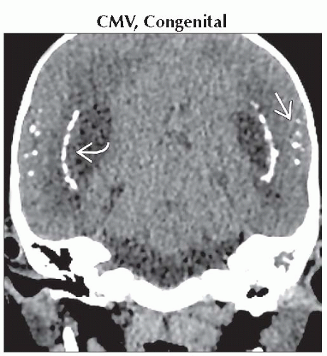 Coronal NECT shows classic findings of TORCH. Note linear periventricular Ca++  with scattered Ca++ foci within cortex with scattered Ca++ foci within cortex  in this deaf child, suggesting prior intrauterine CMV exposure. in this deaf child, suggesting prior intrauterine CMV exposure. |
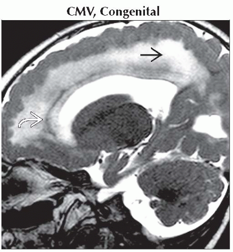 Sagittal T2WI MR shows a thick cortex with small gyri, hyperintense white matter
 , and a thin layer of calcification , and a thin layer of calcification  in the same 18 month old deaf toddler. in the same 18 month old deaf toddler.Related posts:Stay updated, free articles. Join our Telegram channel
Full access? Get Clinical Tree
 Get Clinical Tree app for offline access

|
