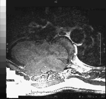
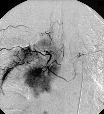
57 A 52-year-old man with new-onset atraumatic back pain was found to have a large T6-T7 vertebral body lesion causing significant cord compression. Following preoperative embolization, he developed progressive right lower extremity weakness. FIGURE 57-1 MRI with a compressive paraspinal mass. Axial magnetic resonance imaging (MRI) of the thoracic spine reveals a right T6 and T7 paraspinal mass involving the vertebral bodies with cord compression (Fig. 57-1). Angiogram with T6 and T7 intercostal injections showed a neovascular stain and a hypervascular mass (Fig. 57-2). FIGURE 57-2 Angiogram demonstrating injection of a hypervascular mass.
Plasmacytoma with Postembolization Swelling
Presentation
Radiologic Findings
Plasmacytoma with Postembolization Swelling
Only gold members can continue reading. Log In or Register to continue


Full access? Get Clinical Tree


