Fig. 1
Pictographs showing representative relative hemorrhage between groups
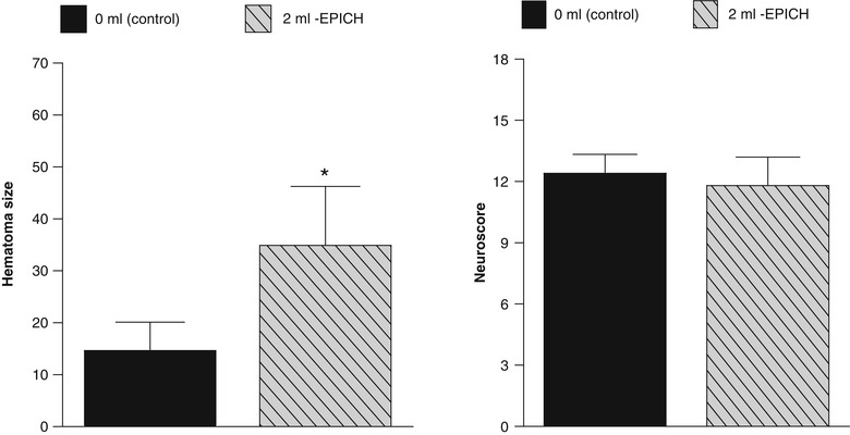
Fig. 2
(Left panel) Hematoma expansion; (Right panel) neurological deficits (sensorimotor skill) measured 24 h following collagenase infusion; SEM, standard error of the mean (asterisk) <0.05 compared with vehicle
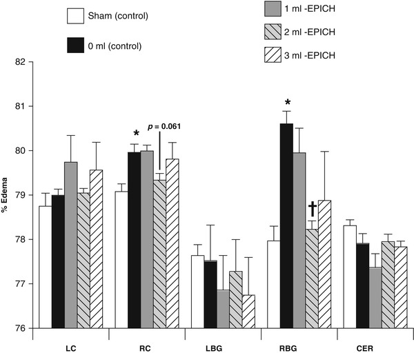
Fig. 3
Percent edema 24 h following collagenase infusion, (asterisk) <0.05 compared to sham; (cross) <0.05 to 0 ml control; SEM standard error of the mean
Conclusion
Translational stroke studies, including animal modeling, are needed to safely integrate basic preclinical scientific principles ahead of clinical application [27–31]. Exsanguination therapy following ICH in rodents, using the traditional phlebotomy approach, may ameliorate the early brain injury (hemorrhage and edema), despite equivocal changes in the short-term neurological functional ability. This study forms the basis to justify further investigation into the preclinical application of this safe and broadly available clinical therapy as a potential therapeutic modality for this devastated patient population. Future studies will need to further delineate the involvement of specific neuroprotective molecules, sympathetic responses, hemodynamic molecules, or neuro-endocrine factors involved in this apparent postconditioning of rat ICH.
Acknowledgment
This study was partially supported by National Institutes of Health grant RO1 NS078755 (Dr Zhang). We wish to thank William Rolland for his handling of a few samples.
Disclosures
None
References
1.
2.
Gao C, Du H, Hua Y, Keep RF, Strahle J, Xi G (2014) Role of red blood cell lysis and iron in hydrocephalus after intraventricular hemorrhage. J Cereb Blood Flow Metab 34:1070–1075PubMedCentralCrossRefPubMed
3.
Zhao J, Chen Z, Xi G, Keep RF, Hua Y (2014) Deferoxamine attenuates acute hydrocephalus after traumatic brain injury in rats. Transl Stroke Res 5:586–594PubMedCentralCrossRefPubMed
4.
Chen Z, Gao C, Hua Y, Keep RF, Muraszko K, Xi G (2011) Role of iron in brain injury after intraventricular hemorrhage. Stroke 42:465–470PubMedCentralCrossRefPubMed
5.
6.
7.
8.
Huang FP, Xi G, Keep RF, Hua Y, Nemoianu A, Hoff JT (2002) Brain edema after experimental intracerebral hemorrhage: role of hemoglobin degradation products. J Neurosurg 96:287–293CrossRefPubMed
Related posts:
 of Behavioral Deficits in Rodents Following Brain Injury Across Species, Gender, and Experimental Model
 Infarction After Aneurysmal Subarachnoid Hemorrhage
 Ganglia Damage in Experimental Subarachnoid Hemorrhage
 Pretreatment Fails to Provide Neuroprotection Following a Surgically Induced Brain Injury Rat Model
 IGF-1 Reduced Rat Pup Germinal Matrix Hemorrhage
 Injection of Noncellular Cerebrospinal Fluid from Subarachnoid Hemorrhage Patient into Rat Ventricles Leads to Ventricular Enlargement and Periventricular Injury
Stay updated, free articles. Join our Telegram channel

Full access? Get Clinical Tree


