Chapter 26 Posterior and Posterolateral Access to the Thoracic Spine
INTRODUCTION
Several surgical approaches have been developed to remove thoracic vertebral lesions from the anterior to posterolateral direction. The choice of surgical approach is decided depending on the disease entity, level of the lesion, laterality, multiplicity, presence of instability, and the necessity of reconstruction. The posterior approach provides less extensive exposure of the vertebral body than the anterior approach, but the involved anatomy is familiar to the spinal surgeons and it can be applied to patients with anesthetic risk.1
Advantages of the posterior approach over the anterior approach are as follows2,3:
With increasing bone removal, from the transpedicular approach to the costotransversectomy, and finally to the lateral extra-cavitary/lateral parascapular approach, the operative trajectory becomes more lateral to better visualize the affected vertebrae.4 In this chapter, overall review of the posterior or posterolateral approach to thoracic spine will be presented with the pertinent anatomical assessment.
MUSCULAR ANATOMY
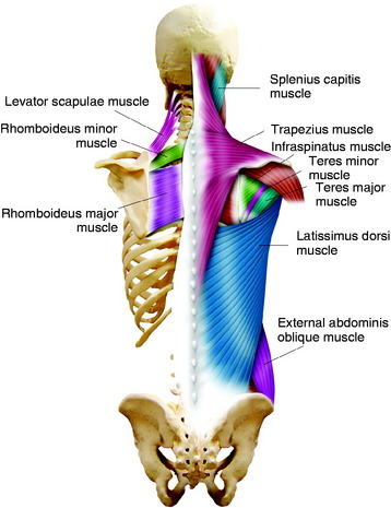
The most superficial muscle of the dorsal spine is the trapezius muscle (Fig. 26-1). The trapezius originates along the external occipital protuberance and each spinous process from C1 to T12. The insertion of the trapezius is the lateral third of the clavicle, the acromion and the scapular spine. This muscle provides the stabilization and abduction of the shoulder. Immediately deep to the trapezius muscle on the upper thoracic level lie the rhomboid major, rhomboid minor, and levator scapulae muscles (see Fig. 26-1). The rhomboid muscles originate from the spinous processes of the cervical and thoracic spine and insert to the ventral edge of the scapula.5 The levator scapulae muscle connects the scapula to the upper cervical vertebrae.
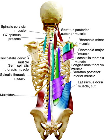
The serratus posterior superior muscle is another muscle that fixes the cervicothoracic junction area spinous processes to the lateral part of rib cage (Fig. 26-2). In the exposure of the cervicothoracic junction, the spinous process insertions of these muscles are taken down as a single group for lateral retraction.6 As these muscles are taken down, the scapula is released from its attachments to the spinous processes and rotates anterolaterally out of the operation field.
On the lower portion of the back, the latissimus dorsi muscle spans over the body. It originates from the spinous processes of the six lower thoracic vertebrae, lumbar and sacral vertebrae, and ilium, inserting onto the humerus (see Fig. 26-1).
The erector spinae muscles are a group of muscles running from the sacrum and iliac crest to the ribs or transverse process of the vertebrae. They have three separated groups: iliocostalis (lateral), longissimus (middle), spinalis (medial) (see Fig. 26-2). The iliocostalis muscle is inserted into the angles of the ribs and into the cervical transverse processes from C4 through C6. The longissimus thoracis muscles are inserted into the thoracic transverse processes and nearby parts of the ribs between T2 and T12. The spinalis muscle is largely aponeurotic and extends from the upper lumbar to the lower cervical spinous processes.
POSTERIOR THORACIC CAGE
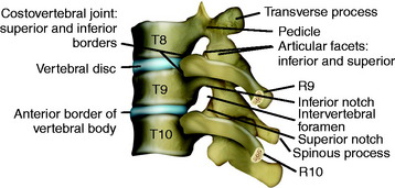
The head of a rib articulates with the adjacent parts of its own vertebral body, the vertebra above, and the intervertebral disc between them (Fig. 26-3).
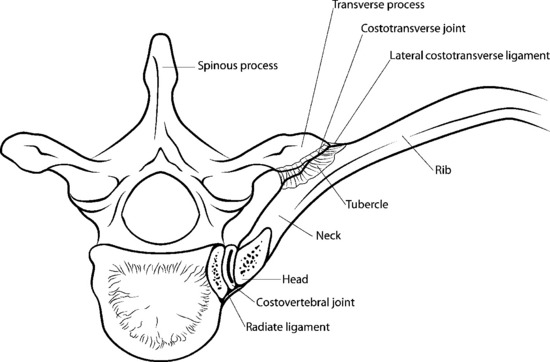
The third synovial joint is a costotransverse joint that is strengthened by superior and lateral costotransverse ligaments (Fig. 26-4). The superior costotransverse ligament joins the neck of the rib to the transverse process immediately above.
The ribs also are attached to one another through the intercostal musculature, which originates medially on each superior rib and inserts laterally on its immediately inferior rib. This strip of muscles contains the intercostal nerve, artery, and vein. Most often, the intercostal vein is most cephalad with the intercostal artery close to it but caudad (Fig. 26-5).
POSTERIOR MEDIASTINAL SPACE AND NEUROVASCULAR STRUCTURE
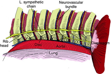
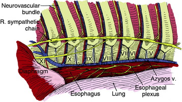
The azygos vein is lateral to the esophagus on the right side, running inferiorly to join the superior vein cava at the fourth interspace. At the point where the azygos vein turns medially, it may receive some branches, which may be divided if necessary (Fig. 26-6).
Stay updated, free articles. Join our Telegram channel

Full access? Get Clinical Tree


