Chapter 22 Posterior Approach to the Cervicothoracic Junction (Pancoast Tumor Surgery)
INTRODUCTION
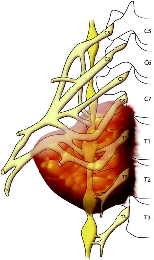
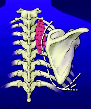
Primary carcinomas arising in the apex of the lung (Pancoast tumors) can extend into the chest wall and the brachial plexus, causing the characteristic Pancoast syndrome (Fig. 22-1). The classic Pancoast syndrome of rib erosion, shoulder pain radiating down the arm, and Horner’s syndrome results from destruction of the first rib extending typically to involve the T1 nerve root and stellate ganglion of the brachial plexus (Fig. 22-2).1
Factors that affect local tumor control and survival are the completeness of tumor resection, TNM (tumor size, node status, metastatic disease) status, and possibly the extent of lung resection.2–4 The completeness of tumor resection is often limited by the degree of spinal and brachial plexus involvement. In the past, spinal involvement was not a surgical indication. However, advanced surgical techniques and magnetic resonance imaging (MRI) for spine and brachial plexus tumors have led to a reassessment of their respectability. Recently, induction chemoradiation protocols have resulted in an improved ability to achieve pathologically complete histological tumor responses, which may ultimately improve the rates of complete resection and long-term survival.
PREOPERATIVE EVALUATION
NEUROLOGICAL EVALUATION
It is important to clinically distinguish T1 nerve root involvement from that of C8 or the lower trunk of the brachial plexus. Pain extending along the ulnar aspect of the forearm to the wrist suggests isolated involvement of the T1 nerve root. These patients do not present with hand intrinsic weakness, and resection does not generally result in significant weakness. Conversely, pain radiating into the hand, especially the fourth and fifth digits, is indicative of involvement of the C8 nerve root or lower trunk of the brachial plexus, and these patients often present with substantial hand intrinsic weakness.5 Resection may result in a flail hand. Both computed tomography (CT) and MRI demonstrate the brachial plexus well, but they often fail to reveal enough anatomical detail to distinguish T1 from C8 or lower trunk involvement. The physical examination is as important as electrodiagnostic tests.
RADIOLOGICAL EVALUATION
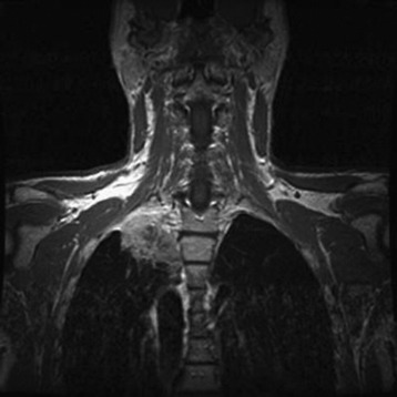
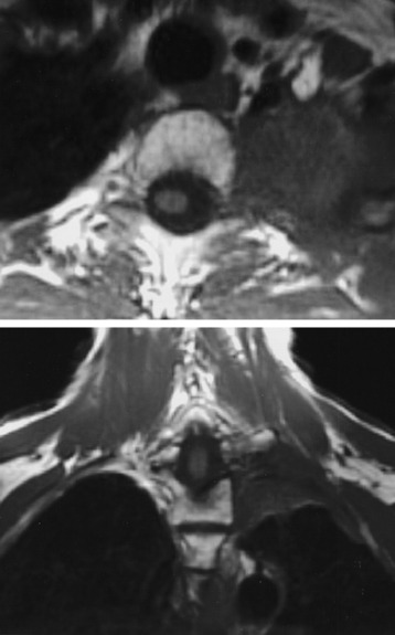
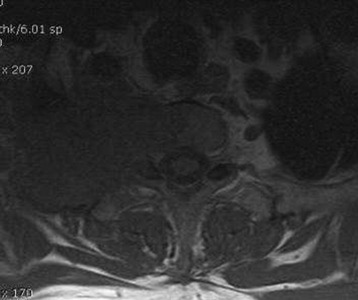
A preoperative MRI classification scheme was devised to assess the degree of spinal element involvement and the type and extent of operation required for tumor resection. MRI findings are divided into four classes based on the degree of involvement of the spinal column and neural structures (Table 22-1).5 Class A tumors involve the vertebral body (VB) periosteum only, and Class B tumors involve the proximal rib head and distal neural foramen without epidural involvement (Fig. 22-3). Class C and D tumors often are not amenable to en bloc resection in which gross-total resection can be achieved. Class C tumors extend through the neural foramen with minimal or no vertebral body involvement but with unilateral epidural compression. Class D tumors involve the vertebral column (vertebral body and/or lamina) with or without epidural compression (Fig. 22-4).
Table 22-1 Radiological evaluation
| Magnetic Resonance Imaging Staging System | |
| Class | Description |
| A | Vertebral body periosteum only |
| B | Extension of tumor into distal neural foramen |
| C | Extension of tumor into proximal neural foramen and/or epidural space |
| D | Vertebral body or posterior element infiltration with our without epidural tumor |
SURGICAL TECHNIQUE
CLASS A AND B LESIONS
Position and Incision
The patient is placed in a lateral decubitus position with the right upper arm abducted.
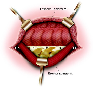
An extended standard posterolateral thoracotomy incision for thoracotomy and posterior midline vertical incision for laminectomy are made (Fig. 22-5). The incision is made midway between the spinous process and medial border of the scapula and extends over the inferior border.
Skin Flap Exposure and Paraspinal Muscle Dissection
After the skin incision is made, the subcutaneous tissue is dissected. The trapezius muscle is incised and reflected along the edge of the subcutaneous tissue. The erector spinae muscle is dissected from the lamina and facet bilaterally from C4 to T8. The dissection plane is made between the spinalis muscle and longissimus muscle in the side the tumor is located. The bilateral erector spinae muscle is retracted to the side that does not involve the tumor (Fig. 22-6). The paraspinal muscles are dissected from the posterior elements of the spine by using a Bovie cautery to expose the transverse processes, pars interarticularis, and unilateral laminae.
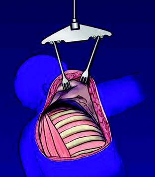
The scapula is elevated from the chest wall by cutting the rhomboid and levator scapulae muscles and is secured with an internal mammary retractor (Figs. 22-7 and 22-8). The chest wall is cut distal to the tumor, most often involving the first through fourth ribs. The chest contents are explored for pulmonary nodes and mediastinal involvement that may preclude resection.

Stay updated, free articles. Join our Telegram channel

Full access? Get Clinical Tree


