Chapter 28 Posterior Approach to the Lumbar Spine
MUSCULAR STRUCTURES FOR POSTERIOR APPROACH IN L-SPINE
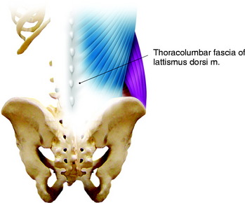
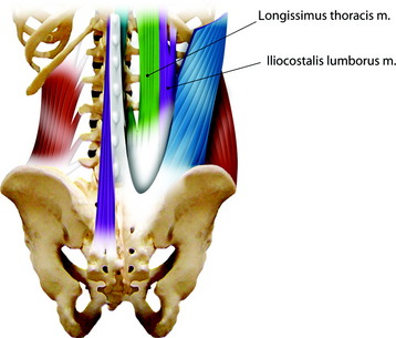
The muscle groups on the dorsal side of the lumbar spine are covered with thoracolumbar fascia. This fascia is the extension of the latissimus dorsi (LD) muscle (Fig. 28-1). Under the LD muscle fascia, the erector spinae muscle groups are located. They are longitudinally arranged with medial, middle, and lateral components (Fig. 28-2). At the upper lumbar level, the serratus posterior inferior muscle is arrayed from the horizontal direction.
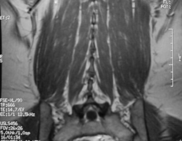
There are cleavage planes between these three groups of muscles. The dissection can be performed more easily between the multifidus muscle and the longissimus muscle than between the longissimus muscle and the iliocostalis muscle (Fig. 28-3).
DEVELOPING THE PLANE DURING MUSCLE DISSECTION
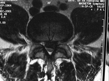
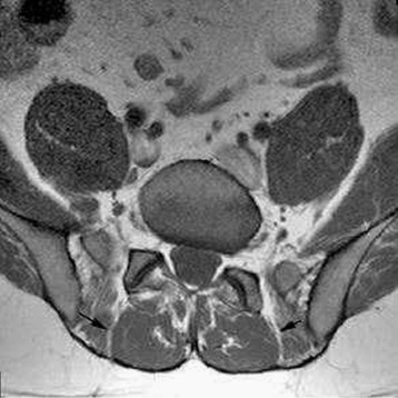
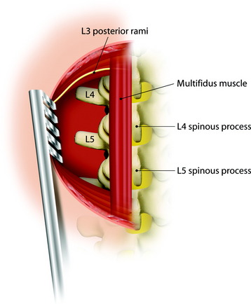
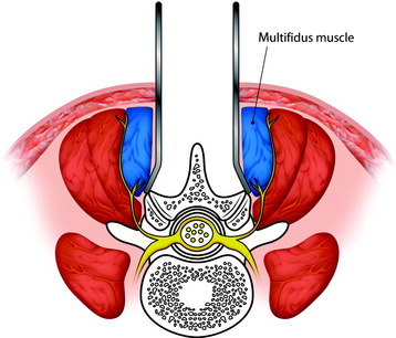
A natural cleavage plane between the multifidus and the longissimus part of the sacrospinalis muscle is present in most of the operated cases. There is a fibrous separation between the two muscular parts. The mean distance between the level of the cleavage plane and the midline was 4 cm (2.4–5.5 cm).1 Small arteries and veins were present, precisely at the level of the cleavage plane. For the dissection between multifidus and longissimus, first, the superficial muscular fascia is opened near the midline, exposing the posterior aspect of the sacrospinalis muscle. Next, the location of the muscular cleft can be found by identifying the perforating vessels leaving the anatomical intermuscular space. The muscular plane can be confirmed with fat tissue. Further dissection leads to the pedicle. The multifidus muscle volume becomes wider from the upper lumbar level to the lumbosacral junction (Figs. 28-4 and 28-5).
NEURAL STRUCTURES IN POSTERIOR APPROACH
The posterior ramus of the spinal nerve root is divided into two branches, medial and lateral (Fig. 28-6). The lateral branch of the posterior ramus of the spinal nerve lies in the fascial plane between longissimus muscle and multifidus muscle (Fig. 28-7). The medial branch of the posterior ramus of the spinal nerve runs into the multifidus muscle belly.
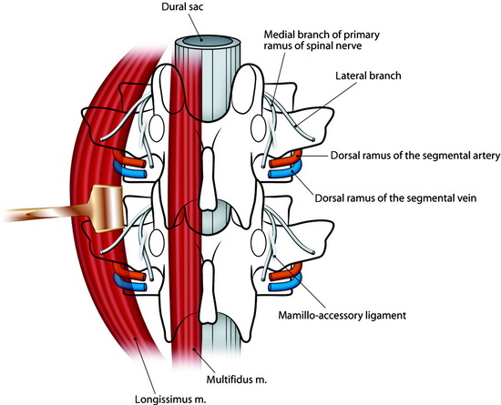
Denervation during a dorsomedian approach to the thoracolumbar spine may cause postoperative atrophy of the deep back muscles. The ensuing instability of the spine with poor clinical results, perhaps as a result of such muscle loss, has been observed in 11.7% of cases.2 Specifically, this complication may be caused by damage to the medial branches of the posterior rami of the spinal nerves during lateral retraction of the muscles.
In the thoracolumbar spine the medial branch of the posterior ramus of the spinal nerve is subject to ligamentous fixation by the strong fibers of the mammillo-accessory ligament, which extends between the mammillary process and the accessory process inferolateral to the superior articular process. When the extent of dissection of the dorsomedian approach to the thoracolumbar spine is enlarged laterally to the articular processes by retracting the paraspinous muscles, the medial branches of the posterior rami of the spinal nerves are endangered (Fig. 28-8). Postoperative pain as well as dynamic instability beyond the corresponding segments could result. To prevent injury to the medial branches of the posterior rami of the spinal nerves, the midline posterior surgical approach to the thoracolumbar spine should not be enlarged laterally to the articular processes.3
Related posts:


Stay updated, free articles. Join our Telegram channel

Full access? Get Clinical Tree


