♦ Preoperative
Operative Planning
- Imaging
- Magnetic resonance imaging (MRI)
- Computed tomography myelogram if MRI is inconclusive
- Flexion/extension x-rays
- Magnetic resonance imaging (MRI)
- Patient counseling regarding surgical risks
- Postoperative pain
- Potential joint instability
- Postoperative pain
Equipment
- Basic spine tray
- High-speed drill (Midas Rex with AM-8 bit)
- One- and 2-mm Kerrison punches
Operating Room Set-up
- Headlight
- Loupes
- Microscope
- Bipolar cautery and Bovie cautery
- Intraoperative x-ray
- Intraoperative fluoroscopy
- Mayfield head holder
Anesthetic Issues
- Consider awake fiberoptic intubation to avoid passive neck extension
- Assess patient’s pulmonary function for ability to tolerate prone position
- Prophylactic intravenous antibiotics (cefazolin 2 g for adults) 30 minutes prior to incision
- Foley catheter for prolonged surgery
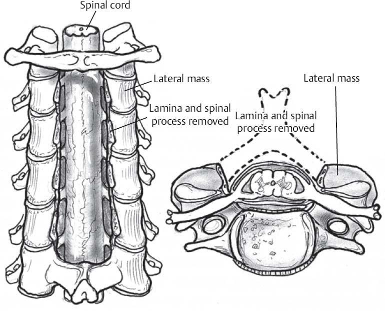
♦ Intraoperative (Fig. 101.1)
Positioning
- Prone position with appropriate padding to prevent pressure neuropathies
- Arms tucked at sides
- Mayfield head holder or tongs with traction to secure head in capital flexion
- Mild reverse Trendelenburg position for venous drainage
- Intraoperative fluoroscopic imaging used to confirm cervical alignment
Planning of Minimal Shave
- Use disposable razor

Fig 101.1 Schematic of posterior cervical decompression.
Only gold members can continue reading. Log In or Register to continue
Stay updated, free articles. Join our Telegram channel

Full access? Get Clinical Tree


