♦ Preoperative
Special Equipment
- Basic spine tray
- High-speed drill with small burr
- Kerrison rongeurs: 2-, 3-, and 4-mm
Operating Room Set-up
- Jackson table (for fusion to preserve sagittal balance) or standard table with Wilson frame (for decompression)
- Headlight
- Loupes
- Microscope (optional)
- Bipolar and Bovie cauteries
- Intraoperative plain x-ray or fluoroscopy
Anesthetic Issues
- Avoid paralytics to maintain peripheral nerve stimulation
- Foley catheter for prolonged cases
- Padded headrest to avoid pressure on face (risk of decubiti) and orbits (risk of retinal ischemia) during prolonged surgery
♦ Intraoperative
Positioning
- Prone position with abdomen free of compression to reduce venous pressure
- Option for Trendelenburg in event of cerebrospinal fluid (CSF) egress from dural perforation
- Arms padded and forward on arm rests
Preparation
- Intravenous antibiotics completed prior to skin incision with coverage of skin flora
- Avoid shaving; utilize electric clippers to thin hair as needed
- Sterile scrub and preparation
- Mark incision with either fluoroscopic guidance, preincision radiograph, or anatomic landmarks
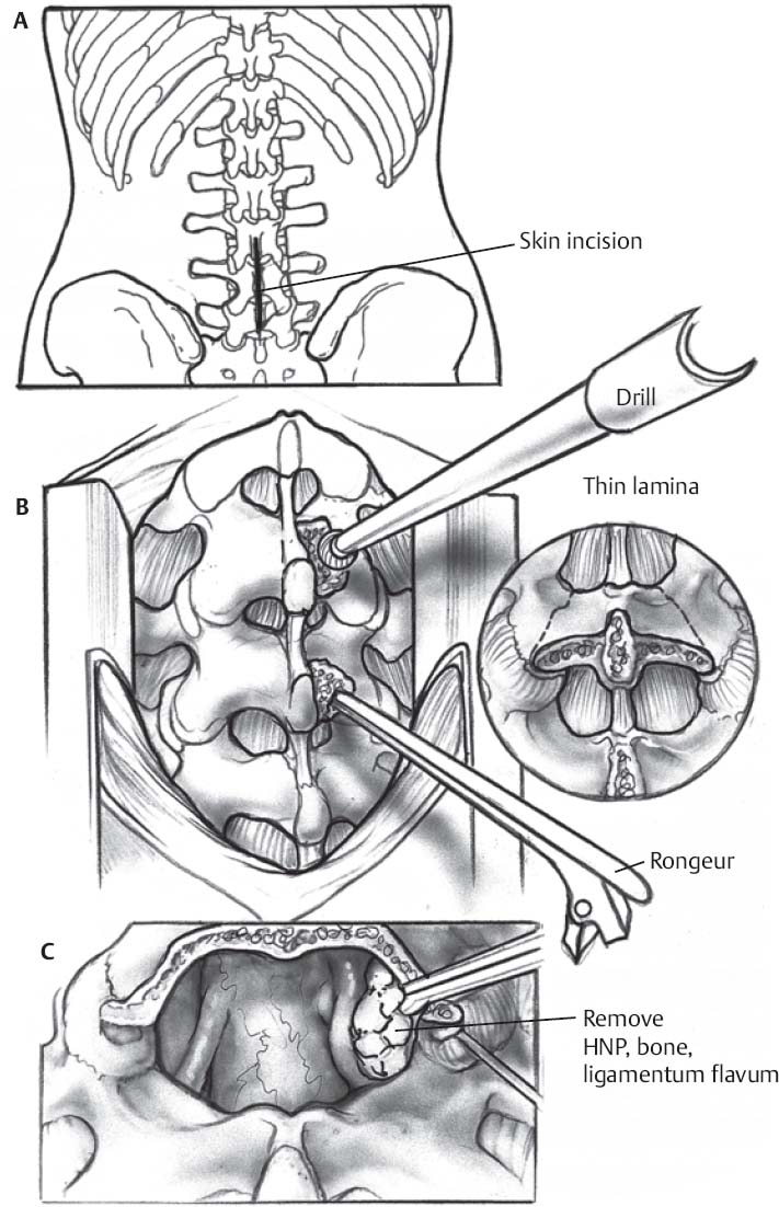
- Exposure(Fig. 119.1A)
- Linear skin incision over spinous processes of interest
- Linear fascial incision over same area with a subperiosteal dissection; the fascial incision may be extended under the skin incision for a broader exposure.
- Intraoperative confirmation of appropriate level with fluoroscopy or plain radiograph
- Bone, ligament, and disc removal (Fig. 119.1B and 119.1C)
- Laminae thinned with Leksell rongeur or high-speed drill
- Larger Kerrison rongeurs used to complete laminectomies with preservation of epidural fat and ligamentum flavum to protect dura
- Elevation of ligamentum flavum and epidural fat to decompress thecal sac
- Careful dissection of ligamentum flavum away from lateral dura
- Small Kerrison rongeurs to undercut facet osteophytes and hypertrophied ligamentum flavum
- Bone and ligament removal continues until surgeon is “flush” with medial pedicle
- Nerve roots may be retracted to accomplish discectomy
Closure
- Valsalva maneuver to ensure no occult dural perforations
- Gelfoam (optional) for epidural hemostasis
- Hemovac drain (optional)
- Absorbable sutures in fascia and subcutaneous space
- Nonabsorbable sutures or staples on skin
♦ Postoperative
- Early mobilization to avoid deep vein thrombosis
- May be discharged next day or same day (after 6 hours) if only single level decompressed
- Analgesics and muscle relaxants as needed

Only gold members can continue reading. Log In or Register to continue


