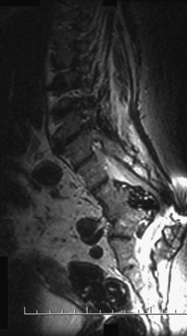
72 A 68-year-old woman had Pott’s disease as a child, and underwent several corrective lumbar procedures. She presented with progressive pain and gait and bowel/bladder dysfunction. Preoperatively, spinal magnetic resonance imaging (MRI) showed severe compression of the dural sac with an abnormal kyphoscoliosis at L2-L3 by a mass likely derived from her previous granulomatous inflammation (Figs. 72-1 and 72-2). In addition, her lumbosacral spine is significantly spondylotic with a previous fusion at L4-L5 noted. Postinfectious deformity Using a bilateral transpedicular approach and redo laminectomy, a ventral extradural mass was encountered. This consisted of calcified, caseating material (Fig. 72-3). This was removed piecemeal to restore the spinal canal. No fusion was needed, because she was autofused from her previous disease. The most common site of extrapulmonary involvement of tuberculosis (TB) is the thoracolumbar spine. TB of the spine is called Pott’s disease, and the infection first affects the vertebral body and then spreads to the disc space. Specifically, the anterior aspect of the vertebral body adjacent to the subchondral plate is the initial spinal area affected. Although relatively uncommon in the modern Western world, Pott’s disease can cause a painful and progressive deformity. HIV-positive and immunocompromised patients are at higher risk. Most of these patients autofuse from the pathology and are left with neurologic deficit.
Pott’s Disease
Presentation
Radiologic Findings
Diagnosis
Treatment
Discussion

Pott’s Disease
Only gold members can continue reading. Log In or Register to continue

Full access? Get Clinical Tree


