Fig. 1
The sequential paired-association task with an asymmetric reward schedule. (a) Two associative sequences (A1→B1→C1 and A2→B2→C2) learned by the monkey. (b) Schematic illustration of time events in a sequential paired-association trial (SPAT). The monkey makes a choice by a saccadic eye movement, as indicated by small yellow arrows. RT response time. (c) An asymmetric stimulus-reward contingency is introduced in reward instruction trials, and used in the following SPATs in one block
Action potentials of single neurons were recorded extracellularly with tungsten electrodes (FHC, Bowdoinham, ME, 0.8–1.5 MΩ) from the LPFC and striatum of the monkeys. We further recorded the activity of a neuron using the new stimuli if its activity was modulated by reward using the old stimuli (A1 and A2). Once a pair of new stimuli was tested with one neuron, it could no longer be used for another neuron. We analyzed the activity of each neuron in two time periods prior to the second cues by a two-way ANOVA. The “first cue period” occurred within 100–500 ms after the first cue onset and the “early delay period” occurred within 500–900 ms after the first cue onset.
3 Results
We recorded the activity of 546 neurons from the LPFC, and the activity of 366 neurons from the striatum of the monkeys while they performed the reward-instructed SPATs with old stimuli (the first cues: A1 and A2). The activity of each neuron was analyzed using a two-way ANOVA: (stimulus (A1 or A2) vs. reward (large or small), P < 0.01) in the first cue and early delay periods, respectively. In this study, we focused on reward neurons that showed only a significant main effect of reward to illustrate how reward information was processed in the LPFC and striatum independently of stimulus properties.
In the LPFC we found 92 and 63 reward neurons in the first cue and early delay period, respectively. There were 113 and 44 striatal reward neurons in the first cue and early delay periods. The proportion of reward neurons in the LPFC was significantly lower than that in the striatum (28.4 % (155/546) in the LPFC; 42.9 % (157/366) in the striatum, χ 2–test, P < 0.01). Within these reward neurons, the activities of 106 LPFC and 100 striatal reward neurons were further recorded using the new stimuli. In the later analysis, we focused on these reward neurons whose activity was recorded using both the old and new stimuli.
We found that the majority of the 106 reward neurons in the LPFC (93/106, 87.7 %) also showed reward-type activity to the new stimuli in the first cue and/or in the early delay periods. Within those 100 striatal reward neurons, 95 neurons showed reward-modulated activity to the new stimuli in the first cue and/or in the early delay period (two-way ANOVA (stimulus vs. reward), P < 0.05).
We were interesting in whether the reward neurons in the LPFC and striatum could predict the reward value of the first cue stimulus (particularly for the new stimulus) presented in SPATs just after reward instruction with C1 and C2. We focused on the neural activity in the first SPAT blocks in which the new or old stimuli had been presented for the first time to each recorded neuron (Fig. 2). We found that when an old stimulus was presented as the first cue, both LPFC and striatal reward neurons discriminated the two reward conditions (large and small reward) from the first SPATs (Fig. 2a, c). However, regional differences in response activity were found when the new stimuli were presented as the first cue. LPFC reward neurons were still able to predict the reward value of the new stimuli from the first SPATs after reward instruction (Fig. 2b) despite the fact that the monkeys had never directly learned the new stimulus-reward contingency. In contrast, striatal reward neurons did not distinguish the preferred from non-preferred reward conditions in the first SPAT (Fig. 2d). After experiencing the contingency between reward and the new stimulus in the first SPAT, these neurons subsequently showed significantly differential activity in the two reward conditions from the second SPAT onwards.
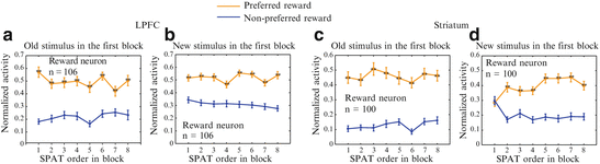
Fig. 2
 Interaction Between Orbitofrontal and Rhinal Cortices Contributing to Reward Seeking Behavior
 Super-Turing Neural Computation
Mind in a Machine: Neural Correlates for Multidimensional Mind Perception
Super-Turing Neural Computation
Mind in a Machine: Neural Correlates for Multidimensional Mind Perception
 State Functions Characterizing the Acquisition of New Motor and Cognitive Skills
 Interactions of Two Individual Arm Robots Using Independent Chaos in Recurrent Neural Networks
 as Bifurcations Shaped Through Sequential Learning


Population activity of LPFC and striatal neurons as a function of SPAT order in blocks. (a) and (b) show the activity of LPFC reward neurons to old stimuli (a) and new stimuli (b). (c) and (d) show the activity of striatal reward neurons to old stimuli (c) and new stimuli (d). The normalized activity was sorted into the preferred reward condition (orange curves) and the non-preferred reward condition (blue curves). Statistical significance was determined by Mann-Whitney U test (** P < 0.01). Error bars indicate the s.e.m
Related posts:
Interaction Between Orbitofrontal and Rhinal Cortices Contributing to Reward Seeking Behavior
Super-Turing Neural Computation
Mind in a Machine: Neural Correlates for Multidimensional Mind Perception
State Functions Characterizing the Acquisition of New Motor and Cognitive Skills
Interactions of Two Individual Arm Robots Using Independent Chaos in Recurrent Neural Networks
Stay updated, free articles. Join our Telegram channel
Full access? Get Clinical Tree
