Preparation for Movement Disorder Surgery

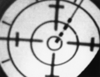
5 Preparation for Surgery


Ron L. Alterman and Michele Tagliati


The elective nature of movement disorder surgery demands strict patient selection criteria and optimal surgical technique to ensure patient safety. This chapter discusses the preparation for surgery from the neurosurgeon’s perspective beginning with the surgical aspects of patient selection and concluding with the various techniques available for anatomical targeting and surgical planning. When appropriate, scientific evidence in support of best practice is presented. Contemporary stereotactic surgeons have at their disposal several techniques that can be used to perform movement disorder surgery. Surgeons must choose an approach that suits their surgical philosophy as well as the strengths of the institution in which they work.


Presurgical Evaluation


Surgery should be considered for any patient with Parkinson disease (PD), essential tremor (ET), or primary torsion dystonia who is cognitively intact and disabled by motor symptoms that are poorly responsive to standard medical regimens. Properly diagnosing and treating movement disorders is no simple matter, and it is the wise neurosurgeon who partners with a movement disorder neurologist who will ensure that the patients are properly diagnosed and medically treated before surgical intervention is offered. This topic is well covered in Chapter 4. Nevertheless, the decision to proceed with surgery and the choice of procedure rest with the surgeon, requiring that the surgeon be well acquainted with the more common movement disorders and can screen for potential problem patients.


Parkinson Disease


Selecting PD patients for deep brain stimulation (DBS) or ablative surgery poses a significant challenge for several reasons. First, PD is highly variable in its clinical presentation and progression.1 Patients suffer from many symptoms, each of which varies in its response to dopaminergic medications and surgery.2,3 Second, care must be taken to identify patients with atypical parkinsonian syndromes because these disorders may have a limited response to surgery.4 Finally, cognitive decline, a prominent feature in elderly patients with advancing PD, must be ruled out because it can be worsened by surgical intervention.57 A well-documented history and physical exam are needed to properly screen the candidates.


A clinical diagnosis of idiopathic PD is supported by the presence of at least two of the following four symptoms: resting tremor, rigidity, bradykinesia, and asymmetrical onset.1 A substantial and sustained response to L-dopa therapy is a key clinical feature confirming the diagnosis and is a strong predictor of the response to subthalamic nucleus (STN) DBS.3 In general, patients who remain responsive to L-dopa but who suffer with progressive motor fluctuations and L-dopa–induced dyskinesiae are the best candidates for STN DBS surgery.3,8


The progression of an individual’s disease and the time frame in which L-dopa-resistant symptoms appear must also be considered. Unusual clinical features observed early in the clinical course suggest the possibility of an atypical parkinsonian syndrome.9 These include (1) prominent postural instability, (2) predominant rigidity or axial symptoms, (3) freezing phenomena (akinesia), (4) hallucinations unrelated to medication, (5) dementia preceding motor symptoms, (6) supranuclear gaze palsy, (7) severe dysautonomia, and (8) early loss of L-dopa response.912 These traits or a history of conditions known to produce parkinsonism (e.g., chronic neuroleptic use, focal brain lesions) strongly suggest a diagnosis of atypical parkinsonism and constitute a relative contraindication to DBS or ablative surgery.


When atypical parkinsonism is suspected on clinical grounds, 18F-fluorodeoxyglucose positron emission tomography (FDG/PET) may be used to determine the proper diagnosis. FDG/PET is the most reliable means of distinguishing idiopathic PD from multiple system atrophy (MSA) or other forms of atypical parkinsonism.6,13 Idiopathic PD is marked by hypermetabolism of the lentiform nucleus on FDG/PET, whereas MSA is indicated by lentiform hypometabolism.13 FDG/PET may also serve as a quantitative predictor of the response to pallidotomy, with the degree of hypermetabolism correlating with postoperative motor improvement.14 The use of metabolic imaging modalities for the evaluation of patients undergoing DBS therapy is currently under investigation.15,16


Visual hallucinations unrelated to medication intake may herald the development of Lewy body dementia.9 Such patients should be excluded from surgical consideration. Patients experiencing medication-induced visual hallucinations may be good surgical candidates, but their surgery should be delayed until they can be stabilized on a medication regimen that does not impair their mental status. We prefer that patients have a clear sensorium for at least 1 month prior to surgery to avoid increased confusion postoperatively.


Dystonia


Dystonia is characterized by sustained, involuntary muscle contractions generating twisting and repetitive movements or abnormal postures.17 Dystonia may be a primary disorder with no obvious underlying cause or may be secondary, caused by any number of etiologies.17 With rare exception (e.g., dopa-responsive dystonia), medical therapies yield limited results.18 Focal dystonias such as spasmodic torticollis or writer’s cramp may be treated effectively with local injections of botulinum toxin,18,19 but this approach is impractical for generalized dystonia, in which many muscles are affected. Multiple reports indicate that DBS can generate profound improvement in patients with primary dystonia, in particular those patients with DYT1 gene mutation.2024 Further study is required to define the optimal clinical indicators for DBS in dystonia.


Essential Tremor


ET is a common movement disorder of the aged, with an estimated prevalence of 0.3 to 5.6% of the general population.25 This is a monosymptomatic ailment characterized by a 4 to 12 Hz postural tremor that is exacerbated with emotional stress and volitional movement.26 The hands and arms are predominantly affected. Head tremor occurs in ~40% of cases, voice tremor in 20%.27 Most patients with ET present with mild, nondisabling tremor.28 Only 10% or fewer develop severe motor disability that interferes with activities of daily living.29 In these cases, medical therapies, including β-blockers and barbiturates, yield limited results. Ablation or DBS in the ventrolateral thalamus yields excellent long-term control of ET, especially tremors of the distal extremities.3032 Unlike PD, in which tremor is the least disabling feature, control of tremor in ET results in significant improvements in functional capabilities.3032


Editor’s Comments


There are important differences between the focus of the neurosurgeon and the neurologist in selecting patients for functional neurosurgery. The neurologist will predominantly consider the appropriateness of the diagnosis and the medical treatment. The neurosurgeon must evaluate the appropriateness of the patient for a surgical procedure but must also overlap with the neurologist in the final screening before surgery. This is especially true for neurosurgeons without the advantage of working in a movement disorder center. Rarely accept the diagnosis without verification by an expert in movement disorders and/or your complete examination.


It all starts with the history. Assume the diagnosis is wrong. It is extremely important that any history which would suggest a different diagnosis be obtained. Although this is certainly possible with a physician extender, the neurosurgeon needs to be involved in this effort to ensure the integrity of the examination diagnosis. Idiopathic PD is a diagnosis of exclusion; therefore, red flags should be noted for patients with a history that includes any of the following: a remitting course, the use of neuroleptic drugs, encephalitis, head injury, stroke, oculogyric crisis, autonomic problems such as hypotension or neurogenic bladder, and rapid progression of disease. Specifically, the history of resting tremor, and a good and sustained response to L-dopa therapy are two of the most critical aspects in defining PD. Tremor is present in ~80% of patients with PD, and its total absence should put up a red flag. Similarly, response to L-dopa is absolutely critical to define idiopathic PD. We do not accept the excuse that the patient is “allergic” to L-dopa because there are multiple agonists that can be used instead. Unfortunately, neither of these is sufficient to exclude other types of diagnoses. The presence of a strong familial tendency in PD suggests that this may not be idiopathic PD, whereas a strong familial history of ET will provide an increased confidence that the ET diagnosis is appropriate. A history of “recreational drug use,” or occupational toxic exposure, whether through agriculture or industry, needs to be carefully reviewed. Dopamine blockers (neuroleptics and antiemetics), dopamine depletors (reserpine and tetrabenazine), and dopamine antagonists (metochlopramide, lithium, α-methyl-dopa, and tricyclic antidepressants) can produce parkinsonian features. Some antiepileptics can cause tremor.


The other aspect of the history that is extremely important for the neurosurgeon is what safety factors may be essential to evaluate preoperatively. Certainly, a list of current medications is essential to prescribing accurate postoperative medication. This is the time to be sure the patient is not on medicine that could cause complications at the time of surgery. Because of the heightened necessity for hemostasis, the history and the list of medications should be scrutinized for any factors or drugs that could delay hemostasis. Specifically, how well does the patient heal following injury, whether or not previous surgeries have been performed without difficulty? Also ask about alternative and over-the-counter medicines because these are frequently not volunteered. The latter is especially important given that multiple medications may contain antiplatelet compounds that the patient may be unaware of. These are listed in Table 5.1.


Any suggestion of a lack of pulmonary or cardiac reserve needs to be carefully evaluated preoperatively by a specialist. This is especially true in PD where cardiac problems may be difficult to detect. A history of sleep disturbance may also suggest that the patient will not be quiet during sedation. Patients with any other type of electrical stimulation devices are generally acceptable. Of most concern are the cardiac pacemakers, and each manufacturer should be contacted to determine compatibility with DBS.33,34 Interference between the two pulse generators (IPG) is possible; appropriate sequencing is necessary to avoid problems. In addition, the IPG device may need to be moved to a different location to avoid programming problems. The presence of a shunt would similarly require special consideration for placement of the IPG, as well as the extension lead. The presence of anticoagulation is a relative contraindication for surgery; however, several patients are on anticoagulants that can easily be discontinued for a week with minimal or no risk. In this situation, we have the internist convert the patient from Coumadin (Bristol-Myers Squibb, Princeton, NJ) to heparin, and then we will reverse it just prior to surgery. Postoperatively, we wait from 3 to 7 days before restarting the Coumadin to ensure adequate homeostasis in the immediate postoperative period. Patients must be forewarned that they are at risk while off medication and the presence of a mechanical device their risk of hemorrhage may be increased postoperatively.


Table 5.1 Medication to Discontinue Two Weeks before Surgery
Aspirin and aspirin-containing products: Alka-Seltzer, Anacin, Ascriptin, Bufferin, Darvon, Compound 65, Ecotrin, Fiorinal, Goody’s Powder, Norgesic, Percodan, Soma Compound, Trilisate
Nonsteroidal anti-inflammatory drugs (NSAIDS): Advil, Aleve, Anaprox, Anasaid, Arthrotec, Cataflam, Celebrex, Clinoril, Daypro, Dolobid, Feldene, ibuprofen, Indosin, Lodine, Motrin, Naprosyn, Nurin, Relafen, Tolectin, Toradol, Vioxx, Voltaren
Blood-thinning medications: Aggrenox, Agrylin (anagrelide), Coumadin (warfarin), Plavix (clopidogrel), Persantine (dipyridamole), Pletal (cilostazol), ReoPro (abciximab), Ticlid (ticlopidine), Trental (pertoxifylline), Lovenox
Other medications suspected of blood thinning: valproic acid, Imuran
Vitamins or herbal supplements may affect the ability to clot such as vitamin E, ginger, garlic, ginkgo, and feverfew
Pain medications that may be taken prior to procedures include Tylenol (acetaminophen), Vicodin (Lortab or hydrocodone), and Ultram

The general physical examination should evaluate patients as to how readily they can undergo surgery and magnetic resonance imaging (MRI). The presence of severe spondylosis, contractures, and kyphosis may reduce the ability to perform the imaging procedure; special considerations may be necessary, such as general anesthesia or a frameless technique. On neurological examination, the presence of the pyramidal signs, absent or atypical tremor, early onset of postural instability, neuropathy, myoclonus, focal cortical signs, and any type of ocular difficulties are all red flags. By definition, ET is a monosymptomatic disorder, so the presence of any additional symptomatology should raise a red flag as to the appropriateness of the diagnosis. Dystonia is widely variable in terms of onset, location, and symptom complexes. As a result, its diagnosis and treatment are extremely complex. This is best discussed in Chapter 12.


Any aspects of the history or physical examination that have raised red flags should be discussed with the referring neurologist to ensure that everything has been done to make the correct diagnosis. Misdiagnosis occurs even among the best movement disorder specialists. If there is a good working relationship, questioning the diagnosis should not be an affront but a legitimate concern expressed by a colleague who wishes to reexamine the diagnosis. Such concerns should not be trivialized. Patients with L-dopa-responsive MSA can be easily misdiagnosed as an idiopathic PD, and DBS STN surgery is not effective.3537 Patients with other atypical parkinsonisms are also not likely to improve following STN DBS (see Chapter 15).


Although the neurologist may suggest the surgical procedure, the neurosurgeon determines which procedure is best for a particular patient. The hemiparkinson patients do very well with unilateral DBS in the STN or globus pallidus internus (GPi). We have patients in postoperative year 6 who have not needed a second sided surgery. Although in general STN DBS is done for PD, a GPi may be an equally good target (see Chapter 15). The ventralis intermedius nucleus of the thalamus (Vim) is used almost exclusively for ET, but STN can also be used (see Chapter 11). Dystonia is predominantly treated with GPi DBS but can be treated with DBS leads in other sites (see Chapter 12). A GPi DBS works very well opposite a successful pallidotomy. Although concerns have been raised about an STN DBS contralateral to a previous pallidotomy,38 we have not found this to be a significant problem for the management of L-dopa therapy, gait freezing, or falls. Clearly, bilateral STN DBS placement can be performed following unilateral or bilateral pallidotomy as well as unilateral or bilateral thalamotomy.7173


The discussion with the patient should be very frank, and it is at this point that the neurosurgeon can determine the expectations of the patient and the family. Both are critically important to correct preoperatively. The family needs to be queried as to their expectations. Families often expect their loved one to be “cured.” If they are not able to live independently postoperatively, the patient might be placed in the nearest nursing home. A stable support system is essential for patients in the postoperative period. Some patients expect to be cured based on media reports or another patient they have seen with a DBS. If improvement is less than anticipated, patients will be unhappy even in the face of marked improvement. It is frequently advantageous to have patients talk to other patients who have undergone the procedure. There is a general rule that patients will not get better than their best “on” time. This is especially important to tell patients because their anticipation of success may far exceed their symptomatic relief. The patient’s age must also be considered. Older patients simply aren’t going to see the maximum improvement that occurs in the younger patients.42 More important than age, however, is cognitive disability. While most patients do not suffer cognitive decline, the potential is always there especially in older patients and patients with less cognitive function preoperatively. Clear dementia or acute psychiatric disorders should be contraindications for surgery. Although gait and postural instability can improve postoperatively, only when these symptoms are minimal in the on stage will improvement be seen.3,4244 Therefore, if these symptoms are severe in the on stage, seriously consider not operating upon these patients, especially if they are older with gait difficulty or there is some suggestion of diagnosis other than the idiopathic PD.


Patients must also come to grips with the potential for complications. Patients should be fully informed of the complication risks, and if they are intimidated it is advisable not to proceed with the surgery. Patients and families must understand that complications can occur at any time. Even when the surgery is successful there are potential problems with hardware or infection. A fifth of patients may require some type of DBS revision.45 They must also understand that their primary doctor postoperatively will be their movement disorder specialist who will manage the medications and the stimulator parameters. Some neurosurgeons do try to manage both, but we have not seen this to be successful. A neurosurgeon can manage the stimulator and the neurologist can manage medications; however, this succeeds only when there is a limited number of patients requiring such interventions. Physician extenders are increasingly called upon to manage these patients, whether they are in neurosurgery, neurology, or both departments.


A few patients will benefit from surgery but will be afraid of the mechanical device and will therefore elect lesioning. In addition, some patients may qualify for surgery, but they may not be candidates for a mechanical device.45 It must be remembered that DBS is an elective procedure, and there are alternative therapies available. As with any elective surgery, there is always a conflict between being too conservative with the indications and offering the procedure to too few patients that might benefit or being too liberal so as to include some patients that will not benefit from surgery. The procedure offered to the patient should therefore be based on each patient’s individual and unique medical, psychological, and social situation.


Preoperative Preparation


Routine preoperative blood work includes a complete blood count, prothrombin time test, and electrolyte analysis. A chest X-ray and electrocardiogram are performed when indicated. Patients are instructed to discontinue aspirin and vitamin E for at least 2 weeks prior to surgery because these agents may increase the risk of intracerebral hemorrhage.46 Antiplatelet agents and warfarin should be discontinued only with the approval of the patient’s medical practitioner. We have on two occasions performed DBS surgery on patients with Von Willebrand disease, providing factor VIII at the time of surgery. We have also operated on two young PD patients who were HIV+. In both instances the patients exhibited stable disease states with normal lymphocyte counts and no episodes of opportunistic infection. Neither suffered a surgical complication, and both have responded beautifully to STN DBS.


Patients with hypertension should take their antihypertensive medications on the morning of surgery because elevated blood pressure may increase the risk of perioperative hemorrhage, and withholding Sinemet (carbidopa/ L-dopa) (Merck & Co., Inc., Whitehouse Station, NJ) for the surgery often results in rebound hypertension. Although not possible in many institutions, it is our practice to admit PD patients the night before surgery. Withholding dopa-minergic medications facilitates microelectrode localization; however, abrupt withdrawal of these medications can result in fever and myolysis similar to that seen in the malignant neuroleptic syndrome.34 Therefore, we prefer to withdraw the patient’s dopaminergic medications in a controlled setting.


Surgical Preparation


Stereotactic Head Frames and Frame Application


Several stereotactic head frames are available commercially. See Chapter 3 for a detailed discussion of the advantages and disadvantages of each stereotactic system. Interested parties are directed to additional references,47,48 which detail the use of the most common stereotactic frames. Most neurosurgeons already have access to a stereotactic frame, which they use for tumor biopsies and difficult ventricular catheterizations. In most instances these frames are serviceable for performing functional neurosurgical procedures; however, we advise having the frame recalibrated by the manufacturer before employing it for this purpose. The accuracy requirements for functional neurosurgical procedures are far greater than those required for brain biopsy, and the performance of frames can degrade over time.


The frame should be selected for its durability, versatility, and ease of use. Most contemporary frames function on the arc-centered principle, which greatly simplifies the targeting process. The frame should allow targeting from both MRI and computed tomography (CT) and should be compatible with the specific independent targeting software that is chosen. The frame should allow the patient to be positioned as comfortably as possible, especially if one plans to use intraoperative microelectrode recording (MER) which can be time consuming. Finally, some form of reticule system should be available so that one may confirm proper intraoperative positioning of the DBS lead or lesioning electrode (Fig. 5.1).


We employ the Leksell Model G stereotactic head frame (Elekta Instruments, Atlanta, GA) for all of the foregoing reasons. The Leksell frame is lightweight yet durable, is both MRI and CT compatible, and allows the patient to be positioned comfortably with the head elevated, minimizing cerebrospinal fluid (CSF) egress from precoronal burr holes. In the operating room, the Leksell frame allows targeting adjustments to be made easily and permits fluoroscopic confirmation of the electrode position.


Head Frame Application

Frame application is perhaps the most overlooked step in performing functional neurosurgical procedures. Proper alignment of the frame with the patient’s anatomy simplifies targeting adjustments and allows the surgeon to use consistent angles of approach. The ear-bars that are provided with the Leksell frame facilitate frame application by preventing sideward tilt (roll) or axial rotation (yaw) of the frame relative to the patient’s head, while permitting the pitch of the frame to be adjusted easily. It is important to use the ear-bar holes that are closest to the frame’s base ring because this raises the frame relative to the body, thus providing enough clearance between the head and shoulders to accommodate the MRI adapter (Fig. 5.2).


image

Only gold members can continue reading. Log In or Register to continue

Stay updated, free articles. Join our Telegram channel

Aug 5, 2016 | Posted by in NEUROSURGERY | Comments Off on Preparation for Movement Disorder Surgery

Full access? Get Clinical Tree

Get Clinical Tree app for offline access