Fig. 1
Brain water content at 24 h; brain water content increased significantly in the right frontal lobe at 24 h post SBI injury. This increase was not reduced following propofol therapy (p > 0.05)
Propofol Therapy Fails to Reduce Neurobehavioral Deficits Following SBI in Rats
Results of modified Garcia test, vibrissae-elicited forelimb placement, and adhesive bilateral tactile stimulation tests conducted at 24 h after SBI showed that vehicle rats presented with severe neurobehavioral deficits compared with sham rats (Fig. 2). However, pretreatment with low- or high-dose propofol failed to reduce neurobehavioral deficits following SBI at 24 h after injury (p > 0.05).
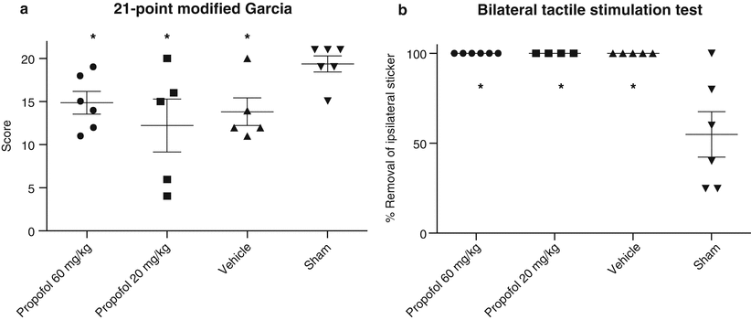
Fig. 2
Neurobehavioral function. Neurobehavioral deficits were present in all vehicle rats compared with sham rats. Following propofol treatment, there was no significant improvement in either (a) the 21-point Garcia or (b) the bilateral tactile stimulation test at 24 h post SBI (p > 0.05)
Discussion
Surgically induced brain injuries can damage viable brain tissue unintentionally by a wide range of methods. The concern with these injuries is that heightened inflammatory response can propagate direct cell death, enhance disruption of the BBB, and subsequently worsen deterioration in neurobehavioral function. Even if patients survive the insult, effective treatment options that improve recovery are limited. Further, no established presurgery therapies are available to help prevent or reduce these collateral damages. In the present study, we investigated the effects of propofol pretreatment therapy on SBI in hopes of demonstrating the potential for this therapy to reduce structural damage at the site of injury and, in doing so, lessen the subsequent sensorimotor behavioral deficits.
Structural brain damage manifesting as cell death and edema following SBI is not an uncommon phenomenon. Previous work in SBI demonstrates massive neuronal degeneration and death in the area of injury [16], including increased expression of proapoptotic factors in cortical neurons and inflammatory cytokines. Study of propofol therapy post-injury using a traumatic brain injury model in rats found that a protective effect can develop through suppression of microglia cell activation and inhibition of a number of inflammatory markers in the ischemic brain [8, 14]. This was echoed in a study that found propofol could reduce lipopolysaccharide-induced inflammatory mediators [7]. Similarly, propofol was shown to reduce cerebral infarct volume by over 30 % using an in vitro model of focal ischemia [1], suggesting preservation of brain structure could occur if inflammatory mediators were attenuated.
Interpretation of our findings is limited by several factors. In our study, vehicle SBI rats had significant damage to their brain structure, as evidenced by a statistically significant increase in brain water content. However propofol pretreatment failed to demonstrate a reduction in brain water content following surgery. The lack of structural preservation was associated with a similar reduction in neurobehavioral deficits following brain injury compared with vehicle treatment, suggesting propofol therapy may not provide neuroprotection as a pretreatment modality in SBI models. However, the timing of treatment could have impacted this result. We chose to inject propofol 30 min prior to the insult to mimic the usual anesthetic course during neurosurgery as closely as possible. However, given the pharmacokinetics of propofol, it is possible there is a need for multiple treatments during and after the injury to demonstrate neuroprotection. To activate and/or amplify targets involved in the plasticity process and improve neurobehavioral deficits, additional propofol doses may be needed after the initial injury. Alternatively, the sheer magnitude of injury caused by this particular established SBI model may be greater than the degree of neuroprotection provided by propofol, thus potentially masking potential impacts on outcome measures.
The results of this study imply that SBI-related morphologic changes in the brain are not affected by propofol pretreatment. Consequently, further studies will be needed to explore the use of propofol therapy in the clinical setting of SBI.
Sources of Funding
This study is partially supported by National Institutes of Health grant NS084921.
Disclosure
All authors attest they have no conflicts of interest to disclose.
References
1.
Adembri C, Venturi L, Tani A, Chiarugi A, Gramigni E, Cozzi A, Pancani T, De Gaudio RA, Pellegrini-Giampietro DE (2006) Neuroprotective effects of propofol in models of cerebral ischemia: inhibition of mitochondrial swelling as a possible mechanism. Anesthesiology 104:80–89CrossRefPubMed
Related posts:
 of Behavioral Deficits in Rodents Following Brain Injury Across Species, Gender, and Experimental Model
 Infarction After Aneurysmal Subarachnoid Hemorrhage
 Volume Determination in Subarachnoid Hemorrhage Using Rats
 Endothelial Growth Factor in Brain Edema Formation After Subarachnoid Hemorrhage
 IGF-1 Reduced Rat Pup Germinal Matrix Hemorrhage
 Injection of Noncellular Cerebrospinal Fluid from Subarachnoid Hemorrhage Patient into Rat Ventricles Leads to Ventricular Enlargement and Periventricular Injury
Stay updated, free articles. Join our Telegram channel

Full access? Get Clinical Tree


