Fig. 1.1
MRI FLAIR and T2* sequences 24 h after onset of left middle cerebral artery infarction. Cerebral oedema is hyperintense on the FLAIR sequence (a star). Deep focal haemorrhagic transformation is visible as a hypointense zone on T2* images b arrow
MRI abnormalities are observed from about the 3rd hour after onset of arterial ischaemia.
T2-Weighted Gradient-Echo
Synonyms: T2-weighted gradient-echo, T2-GE, T2*, T2-star (Fig. 1.1).
T2- and T1-weighted gradient-echo sequences are very sensitive to magnetic susceptibility artifacts.
Methaemoglobin contains iron, which induces artifacts. These sequences are therefore widely used to detect a haemorrhagic component within a zone of infarction or old haemorrhage. Blood has a markedly hypointense signal. This sequence is also useful to demonstrate intravascular thrombus, particularly venous thrombus in the context of cerebral venous thrombosis.
T2 and T1-Weighted Spin-Echo
Synonym: T2 and T1-SE (Fig. 1.2)
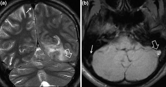
Fig. 1.2
MRI T2 and T1- weighted SE sequences in a female patient with cerebral venous thrombosis. Normal venous blood flow is hypointense on both sequences (a and b single arrows). Absence of venous blood flow in the thrombosed left sigmoid sinus with a hypointense signal on both sequences (a and b hollow arrows)
In addition to demonstrating signs of ischaemia about 3 h after onset of symptoms, as hypointensity on T1 and hyperintensity on T2, these sequences may also visualize arterial thrombus, as blood is markedly hypointense (flow void), while arterial occlusion gives an iso-intense or hyperintense signal (particularly useful for middle cerebral arteries).
TOF or Time of Flight (Fig. 1.3)
TOF is an unenhanced T1-weighted angiography sequence. The vascular signal is favoured over that of surrounding tissues. The tissue signal is saturated and the blood flow signal entering the volume of interest is not saturated, thereby allowing reconstruction of the vessels.
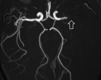
Fig. 1.3
Intracranial MR angiography, TOF sequence in a female patient with left middle cerebral artery infarction. Arterial blood flow in the left middle cerebral artery is interrupted by endoluminal thrombus (hollow arrow)
Gadolinium-Enhanced MR Angiography (Fig. 1.4)
Time-of-flight sequences may present artifacts due to slow or turbulent flow, which is why it is preferable to use contrast-enhanced T1-weighted volume-rendering sequences to explore neck vessels. Signal acquisition is performed during the first pass of the contrast agent bolus in the vessels explored. The contrast agent decreases the T1 relaxation time by a factor of 10, allowing a short repetition time and rapid volume acquisition for three-dimensional reconstructions.
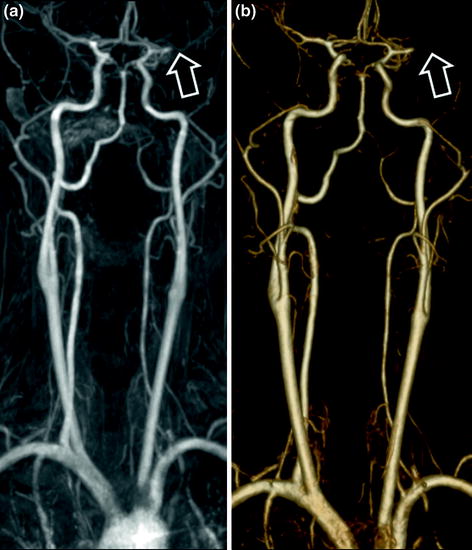
Fig. 1.4
Gadolinium-enhanced cerebral MR angiography (a MIP reconstruction, b 3D volume reconstruction) in a female patient with left middle cerebral artery infarction. Arterial blood flow in the left middle cerebral artery is interrupted by endoluminal thrombus (hollow arrows)
Diffusion-Weighted Imaging
Diffusion-weighted MRI allows exploration of random micro-movements of extracellular water molecules in a medium (Fig. 1.5). These movements depend on the obstacles encountered by water molecules in the medium. In the body, these obstacles essentially consist of proteins, cellular debris or nerve fibres. Free diffusion is said to be isotropic, i.e. water molecules diffuse freely in all directions in a pure medium such as CSF (Fig. 1.6). Isotropic diffusion can be restricted by the presence of an obstacle (protein or cells, etc.), as in the case of an abscess (Fig. 1.7) or a highly cellular lesion (lymphoma) (Fig. 1.8). Finally, diffusion can be anisotropic, for example when water molecules diffuse along nerve fibres (Fig. 1.9). In that case, water molecules no longer move freely in all directions, but preferably diffuse in the direction of the nerve fibres with more limited diffusion in the direction perpendicular to the nerve fibres.
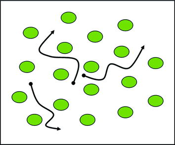
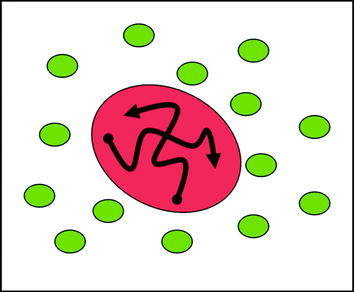
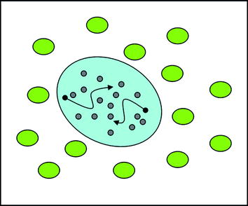
Fig. 1.5
Normal free isotropic diffusion. Water molecules move randomly in all directions
Fig. 1.6
Fluid without cellular debris: increased isotropic diffusion (cyst, tumour necrosis, CSF)
Fig. 1.7


Fluid with cellular debris: decreased isotropic diffusion due to debris (abscess)
Related posts:


Stay updated, free articles. Join our Telegram channel
Full access? Get Clinical Tree
