Fig. 1
The action potential peaks show synchronization for HH nodes grounded with (a) 3 M ohm extracellular resistances at nodes 1–3 compared with (b) zero extracellular resistances, for parallel axons (diameters da = 0.8 μm and db = 2.0 μm)
Figure 2 shows extracellular resistance dependence of the time (ta4 and tb4) of arrival of action potential peaks to the HH node 4 for the axon A of 0.8 μm and the axon B of 2.0 μm diameters, respectively. The effect on the synchronization takes place mainly for the conduction delay for the thick axon gradually depending on the extracellular resistances.
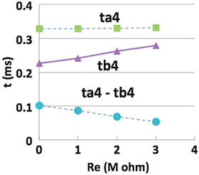
Fig. 2
The action potential peaks reach at node 4 at time ta4 and tb4 with synchronization depending on the extracellular resistances Re for parallel axons of 0.8 μm and 2.0 μm diameters, respectively
5 Discussions and Conclusions
Figure 1 indicates that ephaptic current between the axons causes decrease of the velocity of thick axon resulting the synchronized arrival of the action potentials to the end node. Thin axon, which needs longer time to reach the end node comparing with the thick axon without the ephaptic interaction, causes time-out for synchronization with other neurons. The ephaptic interaction causes sending signals within the time window of the coincident processing on next step. It is effective to maintain functional roles of the thin axon and to provide growth factors of myelination for thin axons in the maturation stages. The extracellular resistance 1 M ohm causes extracellular potential around −25 mV at peak. This high value of extracellular potential is provided by the special structure like that assumed here for the partial encapsulation of adjacent nodes of parallel axons.
The gradual change of synchronization depending on the extracellular resistivity in Fig. 2 provides a possibility of signal conduction regulation by extracellular resistance of glia contribution.
Here we showed that the ephaptic coupling causes (a) signal alignment with adjustment of sending speed. The model also shows several signal processing, i.e., (b) new signal generation and (c) simultaneous signal sending back, and (d) the shifts in the timing of the action potential generation. These processing transfer signals between parallel axons. We did not examine here the three processing, (b), (c) and (d), which will all be reported near future.
Related posts:
 Integrated Neuropsychiatric Assessment System: A Generic Platform for Cognitive Neurodynamics Research
 Dynamic Temporal-Topological Structure of Brain Network Within ADHD
 Mind in a Machine: Neural Correlates for Multidimensional Mind Perception
 State Functions Characterizing the Acquisition of New Motor and Cognitive Skills
 Interactions of Two Individual Arm Robots Using Independent Chaos in Recurrent Neural Networks
Stay updated, free articles. Join our Telegram channel

Full access? Get Clinical Tree


