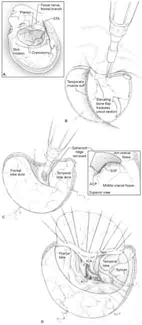
10 Pterional Approach The patient is positioned supine with a bolster under the shoulder ipsilateral to the aneurysm. The head is rotated 15 to 20 degrees away from the side of the aneurysm. The head is extended approximately 20 degrees, allowing gravity to retract the frontal lobe away from the anterior cranial fossa floor and making malar eminence the high point in the surgical field. The head is then lifted above the level of the heart, out of a dependent position. The neck is maintained in a neutral position, avoiding lateral flexion that might close the angle between the shoulder and head, and take away valuable working space. This head position aligns the plane of sylvian fissure vertically, allowing frontal and temporal lobes to fall away naturally to either side as the fissure is split later, like pages in a book that rests on its binding. Retractors become unnecessary during the sylvian fissure dissection. This head position and some lateral rotation of the operating table will adjust for most variability in the plane of the sylvian fissure. A conventional head position with 30 degrees of lateral rotation often leaves the temporal lobe overlying the sylvian fissure and closes the plane, even with full table rotation toward the side of the aneurysm. A curvilinear skin incision begins at the zygomatic arch 1 cm anterior to the tragus and arcs to the midline, just behind the hairline at the widow’s peak (Fig. 10.1A). These two endpoints define the linear fold of the scalp flap, which barely crosses the pterion. Therefore, additional inferior retraction of the scalp flap with “fish hooks” on a Leyla bar (Aesculap; San Francisco, CA) is needed to expose pterion thoroughly. A semicircular incision maximizes the scalp flap. An incision placed too anteriorly along the hairline, having a J– rather than a C-shape, results in a smaller craniotomy because the bone flap conforms to the scalp flap. A foreshortened craniotomy might limit exposure of the posterior sylvian fissure or mobilization of temporal lobe. The scalp is elevated only enough to expose the zygomatic root posterior-inferiorly and the keyhole anteriorly. The superficial fat overlying the temporalis fascia should not be entered because the frontalis branch of the facial nerve lies in this tissue plane and can be injured with additional elevation of the scalp flap. The temporalis muscle is incised from the zygomatic arch to the superior temporal line along the skin incision, then anteriorly to the keyhole, running 1 cm below the superior temporal line. The temporalis is flapped anteriorly, leaving a cuff of fascia and muscle along the superior temporal line to suture the muscle to during closure. The fish hooks are repositioned to retract the temporalis muscle as well as the scalp flap. Patients with large frontal sinuses that will be violated by the craniotomy will require a vascularized pericranial graft for the repair during closure. Head computed tomography (CT) scans or scout films from the angiogram demonstrate the frontal sinus size. It is easier to harvest this pericranial flap during the opening than later during the closure. The depth of the skin incision stops short of the cranium to preserve pericranium, going only through galea and deep connective tissue. The scalp flap is elevated away from the pericranium, opening a white, avascular tissue plane sharply with upward traction on the scalp. The pericranium can be incised well behind the skin incision, extending posteriorly and across midline to enlarge the flap’s size, if necessary. Pericranial flaps elevate cleanly from the bone with blunt dissection and can be preserved during the procedure in moist sponges. Cerebrospinal fluid (CSF) leaks through the frontal sinus are unwanted complications that may require repeat craniotomy, direct repair, and sometimes ventriculoperitoneal shunting. It is far better to prevent this complication than to have to deal with it later when tissues are scarred and the pericranium is compromised. A frontotemporal craniotomy is made using a single temporal bur hole (Fig. 10.1B). The craniotomy follows the temporalis incision posteriorly, then curves anteromedially to the foramen of the supraorbital nerve and inferiorly to the floor of the anterior cranial fossa. This spot is often covered by the fold in the scalp flap, which requires additional retraction during the craniotomy. This seemingly small corner of bone, if it remains, can narrow the outer opening of the operative corridor and limit the maneuverability of instruments held on this side. Dural preservation is particularly important along the inferior bone cut, which is where dura is thinnest. The frontal lobe behind this dura is retracted when pterion is drilled, and tears in this dura can expose brain, leading to injury, swelling, and contusions. Dural integrity is checked by irrigating through the bony cut and shining light directly down on the dura. If there is any suggestion of a dural tear, an additional bur hole can be made at the keyhole, and the dura can be dissected from the inner table of skull. Dural tears are avoided by not crossing pterion with the drill, instead following the floor of the frontal fossa posteriorly. If this dura is torn, intact dura over the orbital roof deep to the tear is elevated and the tear is either repaired primarily with suture or covered with Telfa to protect exposed brain. The pterion is located at the intersection of the frontal bone, the parietal bone, and the greater wing of the sphenoid bone. Pterion lies at the point where the coronal suture intersects with the greater wing, providing an identifiable landmark. Although this point lies on the flat outer table of bone externally, internally the lesser wing of the sphenoid joins the inner table of bone here and is continuous with the orbital surface of the frontal bone, or orbital roof. The inner surface of pterion is a complex three-dimensional structure that prevents its crossing with the foot plate of a drill, and instead requires that it be snapped or cracked to remove the bone flap.
 Position
 Incision
 Extracranial Dissection
 Craniotomy
 Drilling the Pterion

Stay updated, free articles. Join our Telegram channel

Full access? Get Clinical Tree


