Chapter 10 Radial Nerve
Anatomy
Radial Nerve Origin at the Brachial Plexus
• The radial nerve is formed from the posterior divisions of the brachial plexus and is the larger of the two terminal branches of the posterior cord. Receiving contributions from the C5 to T1 spinal nerves, the radial nerve lies posterior to the third portion of the axillary artery at its origin, on the front of the subscapularis, teres major, and latissimus dorsi muscles.
• The fascicles destined for the axillary nerve and the nerve to latissimus dorsi stick to the posterior cord to varying degrees.
• The coracoid process is a reliable landmark at which the radial nerve continues as the main outflow of the posterior cord, posterior to the axillary artery.
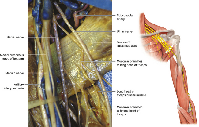
• Close to its origin, the nerve lies on the subscapularis. In its proximal course, the nerve is crossed by the subscapular artery. The size of this stubby vessel varies, and it can be retracted to display the departure of the axillary nerve. The radial nerve lies on the familiar, shiny surface of the latissimus dorsi tendon and then crosses in front of the teres major as the nerve heads, posterior to the subscapular artery, toward the upper end of the spiral groove (Figure 10-1). It courses in front of the long head of the triceps.
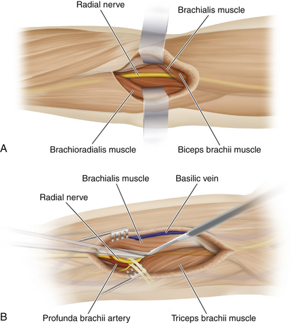
• Also near its origin, the radial nerve gives off a variable number of branches to the triceps. Proximally, the nerve lies behind the brachial artery and in front of the triceps muscle; it deviates from the brachial artery at the point where the nerve winds around the posterior aspect of the humerus from the medial to the lateral side of the arm. The radial nerve passes with the profunda brachii artery through a triangular space bounded by the humerus laterally, the long head of triceps medially, and the teres major superiorly (Figure 10-2).
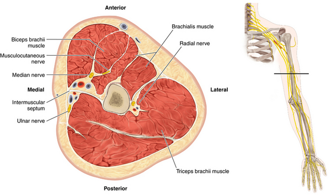
• At the posterior aspect of the humerus, the radial nerve lies in the spiral groove, deep to the long head of the triceps and between the lateral and medial heads. This is the point at which it has the fewest number of fascicles (approximately four or five) in its entire course (Figures 10-3 and 10-4).
• The anatomy (and confusing nomenclature) of the triceps should be clearly understood. The origin of the medial head borders the medial extent of the spiral groove of the humerus. When viewed from behind, the lateral and long heads of the triceps lie side by side, covering the radial nerve, its accompanying profunda brachii artery, and the medial head of the triceps.
• All three heads of the triceps are supplied by the radial nerve, and the surgeon should be aware that these motor branches may leave the radial nerve proximally, as well as in the nerve’s course around the humerus. The anconeus is supplied by a long branch of the radial nerve in the spiral groove. Electromyography of this small muscle may help determine the exact point of pathology along the course of the radial nerve.
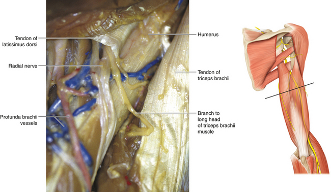
• Throughout its course in the spiral groove, the radial nerve is accompanied by the profunda brachii artery.
• The nerve then runs through the lateral intermuscular septum to gain the flexor compartment of the distal arm (Figure 10-5).

Radial Nerve at the Elbow
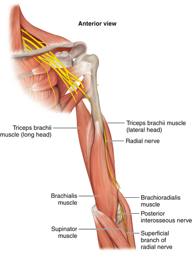
• Lying first in the groove between brachialis and brachioradialis, the radial nerve then descends between the brachialis and the extensor carpi radialis longus to pass in front of the lateral epicondyle into the forearm. The radial nerve gives branches to the brachialis from its medial aspect. The brachialis receives dual innervation from the musculocutaneous nerve and the radial nerve.
• The radial nerve supplies the brachioradialis and the extensors carpii radialis longus and brevis. Muscular branches to the brachioradialis are given off 2 to 3 cm proximal to the elbow.
• When viewed from the front, the radial nerve is easily found lateral to the humerus. The surgeon’s thumb displaces the brachioradialis laterally while the brachialis is displaced medially with the other thumb. The radial nerve will be found at the bottom of this trough (Figure 10-6).
• In patients who are not obese, the radial nerve can be rolled against the humerus as it exits the spiral groove (see Figure 10-5).
Origin of the Posterior Interosseous Nerve
• Note carefully the origin of the ulnar head of the supinator. The muscle fibers wrap around the posterior aspect of the proximal radius and, after embracing the lateral aspect of the proximal radius, are inserted between the anterior and posterior oblique lines of the radius. The superficial head of the supinator is derived from the distal humerus.
• The radial nerve divides into two terminal branches: the posterior interosseous nerve (PIN), which is the deep branch of the radial nerve, and the superficial sensory radial nerve (Figures 10-7 and 10-8).
• The PIN passes between the superficial and deep laminae of the supinator muscle. The supinator has two heads of origin. The superficial (humeral) head has an upper border of variable consistency (muscular, fibrous, or tendinous—the arcade of Frohse). A number of small arterial branches are found at the point where the nerve enters the tunnel between the two heads of the supinator.
• Whereas the branch of the radial nerve to the brachioradialis characteristically leaves the radial nerve from its lateral side, the motor branches to extensors carpii radialis longus and brevis may leave the radial nerve, the PIN, or the superficial sensory radial nerve (or combinations thereof). The motor branches to the supinator may arise proximal to the arcade of Frohse (Figure 10-9) or in the PIN’s course between the two layers of supinator.
• The nerve exits the supinator tunnel and comes to lie between the superficial and deep extensor muscles on the posterior aspect of the forearm. Characteristically, the nerve breaks into numerous fine branches at this point to supply those individual muscles (Figure 10-10).
• One major component supplies the more superficial layer of muscles (extensor digitorum, extensor digiti minimi, and extensor carpi ulnaris) and the second innervates the deeper muscles (abductor pollicis longus, extensor pollicis longus and brevis, and extensor indicis) (Figures 10-11 and 10-12).


Stay updated, free articles. Join our Telegram channel

Full access? Get Clinical Tree


