Radiculopathies and Motor Neuron Disease


Radiculopathies and Motor Neuron Disease


QUESTIONS


1. The following statements regarding radiculopathy are correct except:


A. Pain and paresthesias radiate in a dermatomal pattern


B. Paraspinal muscle spasm is absent


C. Sensory abnormalities are usually vague


D. Muscle weakness is mild if only one root involved

View Answer

1. (B): In radiculopathy, pain and paresthesia radiate in the distribution of a nerve root. This is usually associated with sensory loss and paraspinal muscle spasm. Associated paraspinal muscle spasm commonly limits the range of motion. Each dermatome overlaps widely with the adjacent dermatome. Consequently, it is very unusual to have a severe or dense sensory disturbance. Similarly there is a wide overlap of myotomes, therefore, muscle weakness is mild if only one nerve root is involved. (Preston and Shapiro 1998, p. 413)


2. The most common nerve root that is affected in a cervical radiculopathy is:


A. C5


B. C6


C. C7


D. C8

View Answer

2. (C): In cervical radiculopathy, C7 is the most common nerve root affected followed by C6 and C8 (see Table 17.1). The pain and paresthesia radiate in the distribution of C7 nerve root (neck shoulder dorsum of forearm and middle finger). As there is a wide overlap of myotomes, even in the case of a severe or complete C7 radiculopathy, the triceps brachii is weak but not paralyzed, retaining some strength from its partial C6 and C8 innervation. Triceps jerk is either decreased or absent in the case of C7 radiculopathy. (Preston and Shapiro 1998, p. 413)


TABLE 17.1 Neurologic Signs and Symptoms with Nerve Root Irritation or Damage from Disc Disease


Root


Disk


Pain


Sensory Findings


Motor Findings


Reflex Changes


C5


C4-5


Neck, shoulder, and anterior arm


Lateral arm


Deltoid, external rotators of arm, forearm flexors


Biceps, brachioradialis


C6


C5-6


Lateral arm and dorsal forearm


Lateral forearm, lateral arm, and first and second digits


Forearm flexion, arm pronation, finger and wrist extension


Biceps, brachioradialis


C7


C6-7


Dorsal forearm


Third and fourth digits


Arm extension, finger and wrist flexors and extensors


Triceps


C8


C7-T1


Medical forearm and hand, fifth digit


Medial forearm and hand, fifth digit


Intrinsic hand muscles


Finger flexor


L4


L3-4


Low back, buttock, anterolateral thigh, anterior leg


Knee and medial leg


Knee extension


Patellar


L5


L4-5


Low back, buttock, lateral thigh, anterolateral calf


Lateral leg, dorsomedial foot, large toe


Thigh adduction, knee flexion and dorsiflexion of foot and toes


None


S1


L5-S1


Low back, buttock, lateral thigh, calf


Lateral foot, sole of foot, small toe


Hip extension, plantar flexion of foot and toes


Achilles


(From Brazis PW, Masdeu JC, Biller J. Localization in clinical neurology, 5th ed. Philadelphia: Lippincott Williams & Wilkins; 2007.)


Figure 17.2. The lumbosacral plexus (From Brazis PW, Masdeu JC, Biller J. Localization in clinical neurology, 5th ed. Philadelphia: Lippincott Williams & Wilkins; 2007.)


3. All statements regarding electromyographic (EMG) evaluation of radiculopathy are correct except:


A. If the lesion is acute the EMG study may be normal


B. If the sensory nerve root is predominantly affected the EMG study will be normal


C. Paraspinal muscles are always abnormal


D. Fibrillations in the paraspinal muscles do not imply radiculopathy

View Answer

3. (C): During the first 10 to 14 days after the onset of an acute radiculopathy, there are no needle EMG abnormalities except for decreased recruitment (see Fig. 17.2). If the sensory nerve root is predominantly affected, the EMG study will be normal. In some cases the paraspinal muscles are completely normal and this happens if there is fascicular sparing of fibers to the dorsal rami or may simply be due to sampling error. Similarly, some patients may not relax during the examination making it difficult to assess the paraspinal muscles. Presence of fibrillations in the paraspinal muscles does not always imply radiculopathy as proximal myopathies, motor neuron disease, botulism, and diabetic polyneuropathy affecting the dorsal rami can cause fibrillations. Similarly patients may have persistent fibrillation in paraspinal muscles after spinal surgery. (Preston and Shapiro 1998, p. 421)


Figure 17.1. Tibial F-wave


4. A 45-year-old female patient presented with lower back pain radiating in the right leg. A nerve conduction study should be conducted on all of the following nerves except:


A. Sural nerve


B. Medial plantar nerve


C. Peroneal nerve


D. Tibial nerve (see Fig. 17.1)

View Answer

4. (B): All the above nerves will help to see if there is radiculopathy while medial plantar nerve will not add much information that is not already obtained by the above nerves.

Her sural sensory latency was normal. Peroneal distal motor latency was 5.5 ms with normal amplitude. Tibial distal motor latency was 5.8 ms with normal amplitude with borderline normal F-wave latency (Fig. 17.1). (Preston and Shapiro 1998, p. 417)


5. The differential diagnosis now consists of all except:


A. Lumbosacral radiculopathy


B. Lumbosacral plexopathy


C. Peroneal neuropathy


D. Tibial neuropathy

View Answer

5. (C): Muscles need to be evaluated with needle EMG to assess if there is tibial neuropathy versus lumbosacral radiculopathy. Peroneal neuropathy can be excluded from differential when peroneal motor response is normal and there is no conduction block seen across fibular head. (Preston and Shapiro 1998, p. 311)


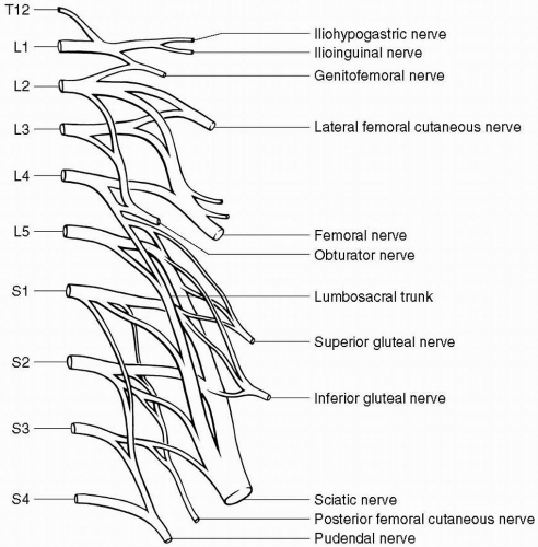
6. A needle EMG of medial gastrocnemius, tibialis anterior, vastus medialis gluteus maximus, and tensor facia lata is performed. The results are as follows:

Medial Gastrocnemius Muscle: Fibrillations 2+, positive sharp 1+, polyphasic motor units 15% of units; recruitment decreased

Tibilis Anterior Muscle: Fibrillations none, positive sharp none, polyphasic motor units none; recruitment normal

Vastus Medialis Muscle: Fibrillations none, positive sharp none, polyphasic motor units none; recruitment normal

Tensor Fascia Lata Muscle: Fibrillations none, positive sharp none, polyphasic motor units none; recruitment normal

Gluteus Maximus Muscle: Fibrillations 1+, positive sharp 1+, polyphasic motor units 15% of units; recruitment decreased

Lumbosacral Paraspinal Muscles: Fibrillations none, positive sharp none, polyphasic motor units none; recruitment normal


The final diagnosis is:


A. L4 radiculopathy


B. L5 radiculopathy


C. S1 radiculopathy


D. Lumbosacral plexopathy

View Answer

Refer to the following clinical scenario for Questions 7 through 11: Clinical Scenario: A 44-year-old right handed man presents to your EMG laboratory for the evaluation of left hand weakness for 3 months.

6. (C): The medial gastrocnemius and gluteus maximus are innervated by S1 root and therefore, S1 radiculopathy is the correct answer. Documenting denervation in proximal and distal muscles innervated by the same root is very helpful in confirming the radiculopathy. Some times presence of denervation only in the distal muscles can be confusing as distal polyneuropathy can give the same pattern. It is important to check proximal as distal muscles. Reinnervation, like denervation occurs in proximal before distal muscles. Sometimes proximal muscles are successfully reinnervated and may show denervation only in distal muscle as is seen in this case where paraspinal muscles are normal. (Preston and Shapiro 1998, p. 420)


7. All of the following tests should be performed except:


A. Left median and ulnar sensory nerve conduction study


B. Left median and ulnar distal motor latency


C. Left median and ulnar F-wave latency


D. Sympathetic skin response

View Answer

7. (D): In the clinical scenario, the patient presents with left hand weakness. Sympathetic skin response will evaluate the small fiber function and not the motor or sensory large fiber functions. Therefore, all the above studies are appropriate except the sympathetic skin response. (Preston and Shapiro 1998, p. 424)

Only gold members can continue reading. Log In or Register to continue

Stay updated, free articles. Join our Telegram channel

Aug 28, 2016 | Posted by in NEUROLOGY | Comments Off on Radiculopathies and Motor Neuron Disease

Full access? Get Clinical Tree

Get Clinical Tree app for offline access