Fig. 19.1
Appearance of lesions in the internal capsule within 1 month and within 24 h after gamma capsulotomy . Right side lesion, 1 month old lesion was caused by 100 Gy, while the left side, 24 h old lesion was caused by 120 Gy. At the time, the lesions appeared the same size, although fainter at 24 h, and the patient’s symptoms were still unchanged [8]
Since the initial efforts of Radiosurgery, functional diseases and specifically for Psychiatry Neurosurgery became important focus of the technique. This occurred until the development of computerized image capable to demonstrate the morphological targets, i.e. tumors and the remarkable response of arteriovenous malformations to single dose of radiation took the attention of the Neurosurgeons interested in the technique. Radiosurgery evolved during the last 20 years of the last century linked to the explosion of the imaging techniques [10]. While dependent on ventriculography, cysternography and angiography until the late 1970s and early 1980s the applications of radiosurgery were largely limited to the pathologies visualized by these techniques. Behavioral surgery applications, as the other functional applications of radiosurgery, were based on principles of functional neurosurgery localization, for example using the anterior commissure (AC) and posterior commissure (PC) seen by ventriculography to guide targeting. Meckel’s cave contrast material injection and cysternography provided visualization of targets such as the trigeminal ganglion in the Mekel’s cave and the acoustic neuroma’s prominence in the cerebello-pontine angle, previously not seen in plain skull radiographs also became focus of the enthusiasts of radiosurgery [11, 12].
19.2 The Inception of the Gamma Knife
Dr. Leksell needing a device capable to treat large number of patients, precise and amenable to the hospital setting recurred to the principle of the cobalt units, then widely used in radiotherapy, to devise the first commercially available, dedicated radiosurgery device. In 1968, Leksell and Larsson developed the first Gamma Knife Unit in Sweden. Larsson was a medical physicist dedicated to develop Gamma Knife and to treat patients with this technique for many decades. The unit was housed in a private setting at the Queen Sophia Hospital (Sophiahemmet) in Stockholm; in 1982 this Unit was transferred to the University of California Los Angeles, being the first Gamma Knife in the USA. This unit was used to treat the first Psychiatric patient with Radiosurgery [8].


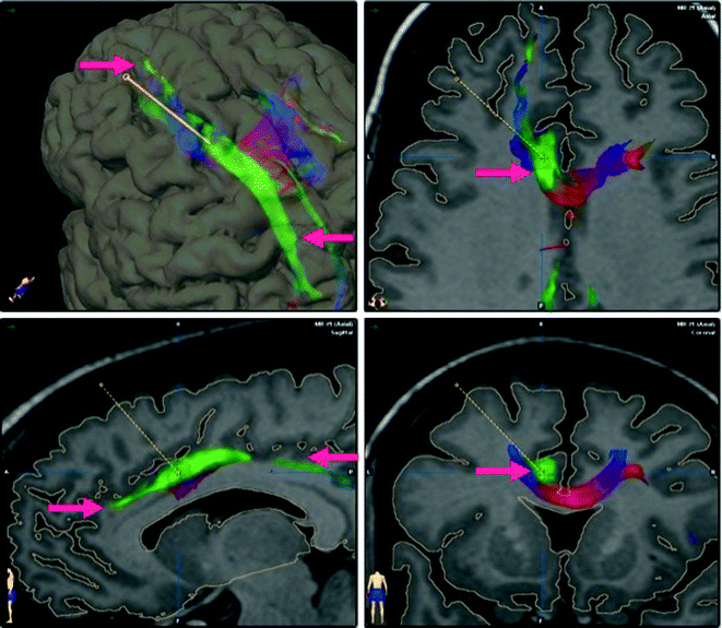
Fig. 19.2
Cingulotomy target sampled with fiber tracking showing spread of fibers mostly to the pre-frontal cortex, high in the frontal lobe a 3D rendering and b axial MRI representation of the cingulate fasciculus spread to the mid-frontal gyrus cortex. Mostly to towards the prefrontal cortex. c Demonstrates the fibers spreading medially in the frontal lobe. d Demonstrates precise location of the sampling in the cingulotomy target
The remarkable results obtained with the Gamma Unit treatment of AVMs, starting in 1972 impressed the neurosurgical community, realizing the potential of the technique as a solution for treatment of these formidable lesions . Angiography provided the visualization of arteriovenous malformations (AVMs), making them the classic application of radiosurgery [13]. Psychosurgery during this period, on the other hand, became highly controlled in the majority of the countries across the world, thanks to the abuse of the trans orbital frontal lobectomy [3]. Therefore losing the center of attention of Neurosurgeons and, specifically of Radiosurgeons that saw in the technique the possibility of treating diseases of easy management and acceptable indications such as AVMs and brain tumors [14]. The technique initially restricted to few institutions and having to provide care for large numbers of patients with structural disease, was not applied in large scale for Psychiatric disorders . Few institutions in the world continue with the work in Psychosurgery , mostly in Spain, USA and Sweden. Careful comparison of radiosurgery with radiofrequency lesions in the brain were carried out at Karolinska, where the main disease treated was OCD with the anterior limb of the internal capsule, the same target was also used for depression during those years [15].
The Gamma Knife evolved to be the only dedicated radiosurgery device for intracranial lesions , competing favorably among neurosurgeons with the various linear accelerator adaptations, when using single dose of radiation, which was dedicated mostly to structural diseases. The appearance of computerized imaging in the 1970s and 1980s amplified the radiosurgery applications, making the demand for dedicated devices for radiosurgery throughout the world [10]. Several models of Gamma Knife represent the evolution of the machine to its state now called commercially Perfexion®. During the evolution of the gamma knife technique lesions in the internal capsule were carried out with searching doses and many times having to adapt to new models, leading to surprises on the size of lesions obtained in the internal capsule, believed to be due to differences in dosimetry between the models of Gamma Knife (Table 19.1).
Table 19.1
Evolution of gamma unit models—technical and economical demands
Gamma Knife U | I. Pioneer: functional neurosurgery (60Co 179 sources) |
II. Initial applications for morphological radiosurgery | |
Gamma Knife B | I. Initial worldwide demand: devices for large-scale treatment and diversity of histology and applications |
II. Economical pressure: replacement of sources at ±7 years interval (60Co 201 sources) became possible | |
Gamma Knife C | I. Computer integration allowing initial efforts of robotization |
II. Computerized treatment plan—replacement of Kula planning—expediting the number of patients treated daily | |
Gamma Knife perfexion | I. Full robotization decreasing possibility of human error |
II. Maximization of collimator interplay for conformality and treatment speed. Replacement of 4 hemispherical helmets of apertures in mm (4, 8, 14, 18) each by one conical helmet with apertures in mm (4, 8, 16) capability, sectors accepting exposure of different number of the 60Co 192 sources available. The GK Perfexion plus brings imaging check capabilities at the time of the treatment |
19.3 Technical Aspects of Radiosurgery
19.3.1 The Energy and Collimation System
60Cobalt decays to 60Ni leading to a half-life of 5.26 years to the cobalt sources powering the GK. The gamma rays resulting from this decay are collimated to the target to achieve the biological effect. Gamma rays of 1.17 and 1.33 MeV are grouped by three different collimation sizes available in the GK Perfexion to automatically take advantage of modulation and shaping capabilities [16]. The previous collimation system of the models U, B and C (Table 19.1), which was dependent on four exchangeable helmets with 4 different sizes of apertures (4, 8, 14, 18 mm) was replaced by a single dynamic conic helmet. This new collimation system is capable of movement throughout three different apertures (4, 8, 16 mm), as well as plugging them strategically to modulate and shape the dose distribution, as desired to optimize the intensity conformity and intensity. The cumbersome process of hoisting the collimators every time that size of the isocenter was changed, serving to delay and bring possible errors to the procedure, is now bypassed in the GK Perfexion [17].
19.4 Flow of Patient Treatment
The psychiatric patient is treated as outpatients after acquisition of the MRI dedicated for the treatment. They are recommended to come to the Gamma Knife department fasting. The day before the procedure they are advised to wash their heads with an antiseptic shampoo. They are advised of the risks of the procedure and sign the informed consent understanding the implications of the radiation, including immediate, delayed and long-lasting effects, as well as the purpose of the procedure, i.e. slow and long lasting effect of radiation in the brain tissue. Therefore the delayed nature of any effect of the procedure in their disease and advised to continue taking their usual medication.
19.5 Placement of the Stereotactic Frame
They are prepared sterile in the forehead and occipital region with topical anesthetic cream followed by injection of 5 cc of mixed Lidocaine/Marcaine and sodium bicarbonate in each stereotactic frame pin site. The frame is applied strategically with the care of including the anterior limb of the internal capsule, i.e. the central part of the brain, AC and PC, central in the stereotactic space. The compatibility of the stereotactic frame placement with all hardware attachments of the Gamma Knife is checked. Measurements of the head surface are acquired with a plastic stick helmet, as well as the measurements of the stereotactic hardware for input in the Gamma Plan for calculation of beam attenuation. The patient is transferred to the CT scan for the stereotactic image acquisition to be merged to the previously obtained MRI. The contour of the patient’s head obtained based on the CT can be used instead of the manual measurements previously obtained to calculate the attenuation of the beams.
19.6 Targeting and Treatment Planning
The target for anterior gamma capsulotomy has been perfected over the years, as well as the dose. Studies are still ongoing to determine whether the most ventral portion of the capsule is most effective or even if there is a lateralization on the brain, which could lead to need of only one side lesion. Now that lesions can be seen, as well as the fibers interrupted by the these lesions can be identified with MRI fiber tracking techniques, understanding may improve on the effects specific lesion localization with consequent optimization of the results (Fig. 19.2).
The initial targets for capsulotomy as described by Mindus et al. [9] was 10 mm in front of the AC, 8 mm above the inter-commissural line and on average 17 mm lateral to the mid-plane. This target was chosen when the first Gamma Unit was available in Stockholm. It was applied a cross-firing of 179 gamma beams precisely collimated with 3 × 5 mm beams. On the basis of experience gained from post-mortem observations on patients subjected to gamma thalamotomy for cancer pain, it was used a central irradiation dose of 160 Gy. The treatment planning now available, the Gamma Plan, takes advantage of full computerized system which can optimize the shape of the lesion, even using different sizes of collimators to reach most ventral portions of the internal capsule in an elongated shape, it has been suggested double isocenters of 4 mm (Fig. 19.3).
Stay updated, free articles. Join our Telegram channel

Full access? Get Clinical Tree


