10 Radiosurgery in Acoustic Neurinomas
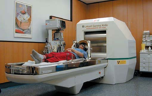
Fig. 10.1 The type B gamma unit (a 201-sourced cobalt-60 gamma knife), which was installed at the Department of Neurosurgery in Vienna in 1992. The radiosurgical unit is independent, although directly connected to the department’s other units and managed by a neurosurgeon.
Since the beginning of stereotactic radiosurgery, the technique has gained increasing importance—on the one hand, as an additional treatment facility after the appearance of a recurrent tumor, but sometimes even as an alternative choice of treatment in elderly patients with concomitant medical problems, or in patients who decline open surgery. Radiosurgery involves precise closed-skull destruction of an intracranial target by applying ionizing beams of radiation. The technique originated in Sweden in 1968, and the first reported treatment of an acoustic neurinoma using the gamma knife was in 1969.
Several recent publications on experience with the techniques involved have reported acceptable results. The avoidance of surgical major problems such as bleeding, infection, and cerebral fluid leakage makes the procedure a comfortable one for the patient, and in the majority of cases, patients can be released from hospital within 24 hours of treatment. In contrast to open surgery, the aim of radiosurgery is not to remove the tumor, but rather to control the tumor’s size, with a minimum of sensory deficits. Due to the morbidity of cranial nerve deficits, the dosage has been reduced to 12–14 Gy in recent years.
However, radiosurgery is not without complications. Cranial nerve deficits, hearing loss, and hydrocephalus have been frequently reported in the literature, particularly in large tumors. Moreover, 15–20 % of the tumors continue to grow after radiosurgery, and there have so far been no publications reporting complete tumor removal and fully preserved function that could compare with the results in microsurgery.
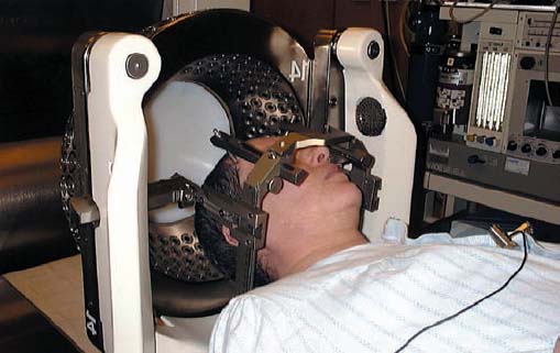
Fig. 10.2 Patient positioning with the stereotactic frame in the outer collimator system. After the Leksell frame has been fixed under local anesthesia to the patient’s skull, an MRI scan is performed (T2-weighted, axial, 2 mm slice thickness; and T1-weighted with contrast, axial and coronal, 3 mm slice thickness). In addition, computed tomography is performed in all patients for potential correction of imaging failures in MRI scans relative to bone–soft-tissue delineation in the internal auditory canal.


Stay updated, free articles. Join our Telegram channel

Full access? Get Clinical Tree


