Fig. 1
Mortality rate in this study (a). Grading score of subarachnoid hemorrhage (SAH) results at 24 and 72 h (B). Wet-to-dry weight ratio at 24 h after SAH (c). In the wet-to-dry weight ratio at 24 h, the low- and middle-dose treatment ameliorated the lung edema compared with the vehicle group. Administration of both the middle dose of JWH133 and the CB2R antagonist, SR144528, reversed the protective effect of JWH133 (SR + JWH(1.0)). *: p < 0.01 vs. vehicle, †: p < 0.01 vs. SR + JWH(1.0), ‡: p < 0.05 vs. vehicle, §: p < 0.05 vs. SR + JWH(1.0) (b). In the wet-to-dry weight ratio at 72 h, no differences could be seen among the groups (d). Values are expressed as median and 25th to 75th percentiles (b) and mean ± SD (c, d)
MPO is a major component of azurophilic granules of neutrophils and polymorphonuclear leukocytes. The levels of MPO were significantly higher in vehicle compared with sham and JWH groups (Fig. 2a). The levels of JAM-A were significantly lower in vehicle compared with sham and JWH groups (Fig. 2b). The levels of MPO or JAM-A in SR-JWH showed the reverse tendency compared with that in JWH group. We also performed immunofluorescent staining of MPO after cardiac perfusion to further determine MPO existence in the alveolar and interstitial spaces, not in the vessels (Fig. 3). Sham did not demonstrate MPO positive cells. On the other hand, vehicle showed several MPO-positive cells. JWH133 treatment reduced the number of MPO-positive cells in the lung.
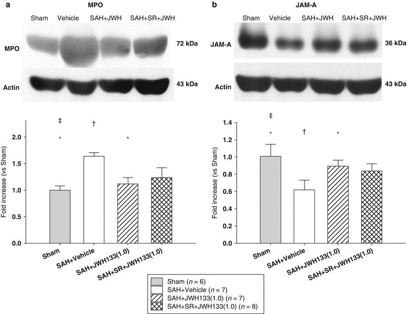
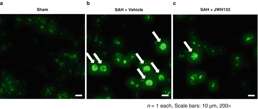
Fig. 2
Representative Western blots and quantitative analysis after SAH. Myeloperoxidase (MPO) (a) and junctional adhesion molecule – A (JAM-A) (b) in the right lung lobes were evaluated 24 h after SAH. Expression levels of each protein in Western blot are expressed as a ratio of actin levels for normalization. Values are mean ± SD. *: p < 0.01 vs. vehicle, †: p < 0.01 vs. SR + JWH(1.0), ‡: p < 0.05 vs. SR + JWH(1.0)
Fig. 3
Representative pictures of immunofluorescent staining with MPO. FITC signals (arrows) indicate MPO-positive cells expressing infiltrated neutrophils in the alveolar and interstitial spaces in the left lung tissues. Sham (a), SAH + vehicle (b), and SAH + the middle dose of JWH133 treatment (c)
Discussion
In this study, we demonstrated that SAH-triggered NPE was ameliorated by JWH133. JWH133 attenuated NPE induced lung edema and neutrophil infiltration into the lungs, which was associated with increased JAM-A expression in the lung 24 h after SAH. However, NPE no longer occurred at 72 h, suggesting that NPE is not frequent after 72 h. To the best of our knowledge, this is the first report to describe the protective effects of CB2R agonist on NPE immediately after SAH.
One putative mechanism of NPE is that increased intracranial pressure causes excessive release of catecholamine, resulting in high permeability. Meanwhile, SAH can cause a systemic inflammatory response, which activates capillary leakage permeability. Norepinephrine also causes activation of cytokines and inflammatory reactions associated with recruitment of neutrophils [15], which result in NPE by damaging the alveolar capillary barrier [11].
In this study, the exposure of rats to SAH increased MPO activity and lung edema in the tissue, indicating the infiltration of polymorphonuclear neutrophils and the development of increased capillary permeability. Immunohistochemical assessment also confirmed that leukocytes infiltrated from vessels substantially in the model of SAH. JAM is a small immunoglobulin family and one of the main constituents of TJs that are important for lung defense [14]. JAM-A is expressed by endothelial and epithelial cells and can mediate leukocyte diapedesis; however, reported roles of JAM-A in leukocyte mediation are not consistent [8]. In the context of acute NPE after SAH, we demonstrated that JAM-A was damaged in the lung, which is consistent with damage in the brain at the same time point (24 h) of SAH [1]. Finally, CB2R activation may represent a potent target for the future development of novel therapies in NPE after SAH through attenuation of TJ disruption and preventing neutrophil infiltration. Further studies are necessary for clinical translation.
Related posts:
 of Behavioral Deficits in Rodents Following Brain Injury Across Species, Gender, and Experimental Model
 Infarction After Aneurysmal Subarachnoid Hemorrhage
 Volume Determination in Subarachnoid Hemorrhage Using Rats
 Pretreatment Fails to Provide Neuroprotection Following a Surgically Induced Brain Injury Rat Model
 IGF-1 Reduced Rat Pup Germinal Matrix Hemorrhage
 Injection of Noncellular Cerebrospinal Fluid from Subarachnoid Hemorrhage Patient into Rat Ventricles Leads to Ventricular Enlargement and Periventricular Injury
Stay updated, free articles. Join our Telegram channel

Full access? Get Clinical Tree


