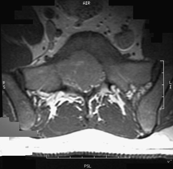
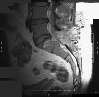
61 A 29-year-old man initially presented with low back pain and bilateral radiculopathies. He had a posterior resection of an S1 lesion followed by 48 Gy external beam radiation. A few months later, he had recurrent bilateral S1 radiculopathies without bowel, bladder, or sexual dysfunction. On examination, he had decreased sensation in the left S1–S3 dermatomes, absent left ankle jerk, and left plantar flexion weakness. FIGURE 61-1 Axial MRI scan showing a destructive sacral lesion impinging on the canal. On magnetic resonance imaging (MRI) of the lumbosacral spine, there is a bony destructive lesion involving the left S1 segment, extending into the canal. It displaces the thecal sac and impinges on the S1 nerve root (Figs. 61-1 and 61-2).
Recurrent Giant Cell Tumor
Presentation
Radiologic Findings
Recurrent Giant Cell Tumor
Only gold members can continue reading. Log In or Register to continue


Full access? Get Clinical Tree


