Reason
Total #
Subclass
#
Trauma
195
RTA: Boda-Boda 135, Auto 19.
154
Normal
14
Assault
28
Fall
13
CVA
46
Ischemic
15
Hemorrhagic:
28
(Subdural
10
Epidural
2
Intracranial
13
Interventricular)
3
Normal
3
Exclude SOL (space occupying lesion)
33
Head symptoms
24
Ear
5
Mouth
1
Nasal
3
Optic
14
Skull
1
Tumor
19
Neural
5
Non-neural
14
Seizure
12
Toxoplasmosis/HIV
12
Headache
12
Spinal symptoms
10
Facial injury
10
Trauma
5
Palsy
4
Tumor
1
Dementia
8
Congenital abnormalities
7
Follow up
5
Psychiatric disorders
4
Encephalitis
3
LOC (loss of consciousness)
3
Grand total
403
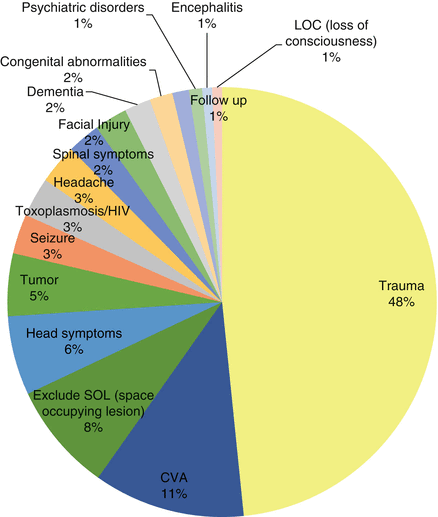
Fig. 14.1
Pie Chart demonstrating
What Where the Most Common Indications That Required a Head-Neck CT-scan? (Fig. 14.2)
We found the majority of the indications were due to Trauma which involved 48 % of the cases followed by Cerebrovascular accidents, CVAs, as the next most common indications affecting 11 % of the cases.
Head Trauma (Fig. 14.2)
We identified a total of 197 patients that were imaged due to a traumatic incident and in this category abnormalities were noted in CT-scans from 154 cases of road traffic accidents, RTAs, 28 cases of Assault and 15 cases due to a fall.
Road Traffic Accidents or RTAs, (Fig. 14.2a), formed 77 % of the total cases of head trauma. There were 154 cases of trauma due to RTA with 10 % (14 cases), demonstrating no abnormalities in their CT head scans. The vehicles that were involved in these accidents were in 12 % (19 of the cases) due to accidents in automobiles or trucks while the majority, 88 % (135 patients) were due to accidents on the small ubiquitous motor bike taxis called “boda-boda” in Uganda (Table 14.1).
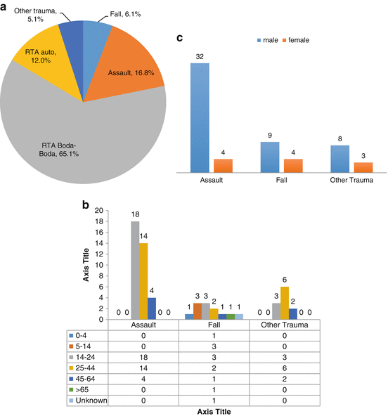
Fig. 14.2
(a) Trauma subclasses; (b) gender distribution of trauma subclasses; (c) age distribution of trauma subclasses
Traumatic Brain Injuries (TBI)
Of the 154 patients with trauma from RTA, 89 % had traumatic brain injuries, TBI, while 61 % of the imaged patients due to assaults and falls had TBI. In the RTA sub-category, males were more commonly affected with 119 males vs. 34 females (Fig. 14.3a) and the most common ages of the affected were in the 14–44 year age bracket (Fig. 14.3b).
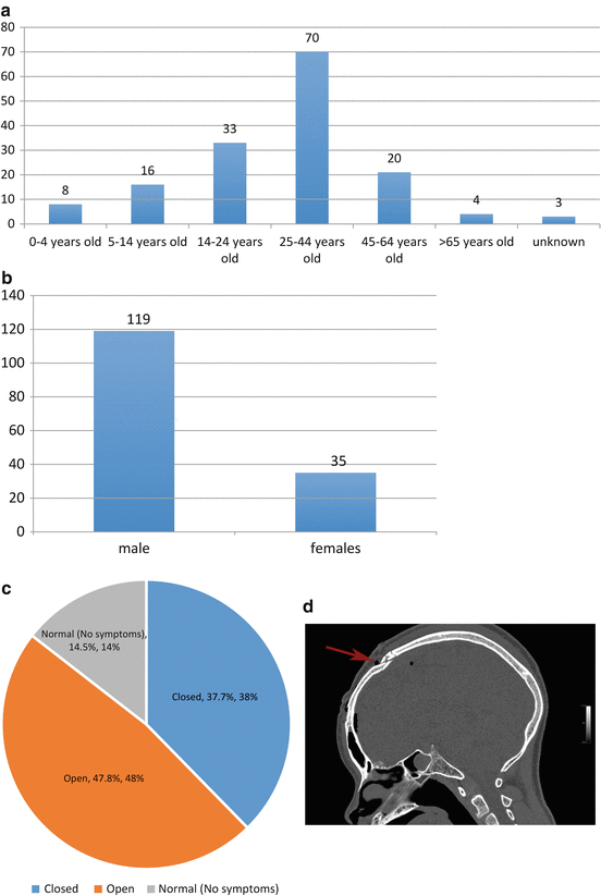
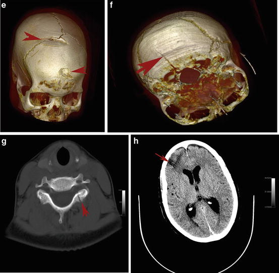
Fig. 14.3
(a) RTA head injury age demographic by age; (b) RTA head injury gender demographic; (c) RTA head injuries open head injuries vs. closed head injuries; (d) CT, sagittal image, fracture of frontal bone, red arrows, after RTA, male age 24 with headaches, red arrow; (e) 3D reconstruction of skull shown in (d) with fracture of frontal bone, red arrows, in a male age 24 (OsiriX imaging Software version 5.9); (f) RTA 3D reconstruction of a skull with a fracture, red arrow, of left occipital bone in male age 14 with headaches (OsiriX imaging Software version 5.9); (g) RTA CT fracture of left lamina, red arrow, in cervical vertebrae C2 male age 19 with pain and some muscular symptoms; (h) RTA. CT image demonstrating the effects of an intracerebral contusion to the right frontal polar cortex due to a RTA in a 64 year old female. The patient demonstrated some changes in personality and upper motor neuron signs on the left side of the body
In Fig. 14.3c in the RTA category we noted that the patients that were imaged due to head injuries consisted of 47.2 % with closed head injuries (CHI), and 37.7 % with open head injuries. All the patients that had either open or closed head injuries had neurological deficits. Of the patients admitted for evaluation of head injuries, 14 % were found to have no abnormalities in their head CT-scan images.
Trauma Due to Either Assault or a Fall (Fig. 14.2a–c)
The other common causes of trauma which led to a patient being imaged included 28 cases of Assaults and this formed 13 % of the cases of trauma, and 13 (6 %) were cases of Falls with 10 % of these cases showing no abnormality in their CT-scan images. These assault and fall injuries were more common in males in 49 cases vs. 11 in females There were another 10 cases listed with 5 of them having brain edema from many sources including infections. The remaining 5 cases were from other forms of unspecified causes and these were all without any abnormalities in their CT-scan images.
Imagery of Skull Fractures from RTA
Figure 14.3d–h, are examples of skull fractures seen in several of the patients caused by RTAs.
Related posts:
 A Case Report of Mania and Glioma
Environmental Toxins as Causes of Brain Degeneration in Sub-Saharan Africa
A Case of Alzheimer’s Dementia in Uganda
Vitamin Deficiencies and Neuropsychiatric Disorders in Sub-Saharan Africa
A Case Report of Mania and Glioma
Environmental Toxins as Causes of Brain Degeneration in Sub-Saharan Africa
A Case of Alzheimer’s Dementia in Uganda
Vitamin Deficiencies and Neuropsychiatric Disorders in Sub-Saharan Africa
 Caring for the Elderly with Dementia in Africa
 CT Findings in the Brain of Adult Patients with HIV/AIDS
Stay updated, free articles. Join our Telegram channel

Full access? Get Clinical Tree


