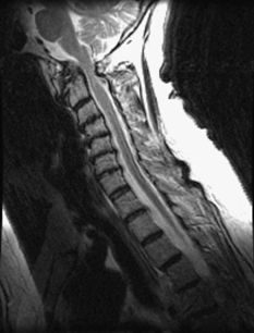
20 A 62-year-old woman with a history of rheumatoid arthritis developed progressive hoarseness and difficulty with swallowing. On examination she had lower cranial nerve palsies and hyperreflexia. Sagittal T2-weighted magnetic resonance imaging (MRI) and x-rays of the cervical spine show evidence of atlantoaxial instability, cord compression from a pannus and C1, and cord signal change at C1 (Figs. 20-1 and 20-2). Atlantoaxial instability and compression from rheumatoid changes A cervical decompression, reduction, and fusion from C0 (occiput) to C3 was performed (Fig. 20-3, lateral cervical x-ray). Rheumatoid patients commonly have neck pain, but only about a third have neurologic deficit. Of the cervical spine involvement, atlantoaxial instability is the most common and is due to an erosive synovitis that is part of the rheumatoid disease process. Pannus formation is due to the micromotion that occurs with ligamentous laxity. The patient who is neurologically intact can usually be watched for progressive atlantodental interval increase (over 8 mm) or platybasia before surgery is warranted. But if the patient shows signs of cervicomedullary compression, a surgery to decompress and stabilize is needed. Rheumatoid patients are a high-risk surgical population because of their degree of systemic illness and long-term use of steroids and other anti-inflammatory medication.
Rheumatoid Arthritis with Instability
Presentation
Radiologic Findings
Diagnosis
Treatment
Discussion
Rheumatoid Arthritis with Instability
Only gold members can continue reading. Log In or Register to continue


Full access? Get Clinical Tree


