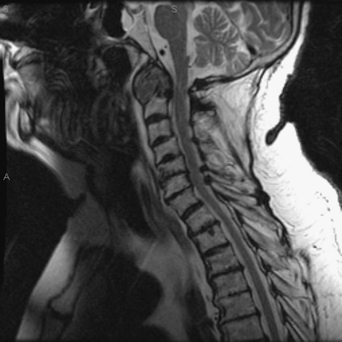
19 An 81-year-old man with a history of rheumatoid arthritis became progressively quadriparetic. He also complained of neck pain. On examination his motor strength was 3/5 in all of his extremities. Sagittal magnetic resonance imaging (MRI) of the cervical spine demonstrates a large, compressive pannus as well as subaxial spondylosis (Fig. 19-1). Pannus Occipitocervical stabilization was performed. Rheumatoid arthritis causes synovial inflammation, ligamentous laxity, and osseous erosion. All of this joint destruction causes micromotion at the occipitoatlantoaxial region, which can result in pannus formation. The pannus itself can compress the cervicomedullary junction, resulting in lower cranial nerve palsies, weakness, and myelopathy.
Rheumatoid Patient with Ventral Cord Compression
Presentation
Radiologic Findings
Diagnosis
Treatment
Discussion

Rheumatoid Patient with Ventral Cord Compression
Only gold members can continue reading. Log In or Register to continue

Full access? Get Clinical Tree


