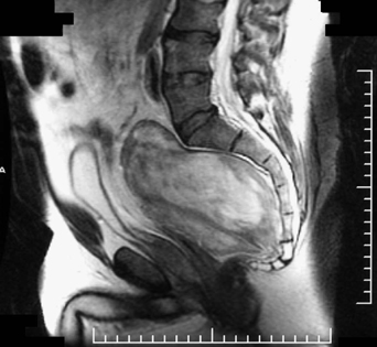
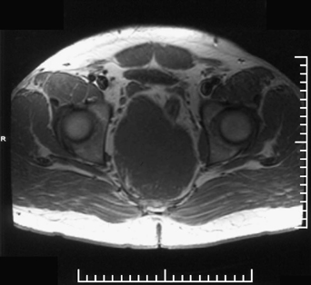
68 A 38-year-old man was troubled by lower extremity radiculopathies. Although he did not have incontinence, urinary frequency with decreased sensation in a right L5 to S3 distribution developed. An urgent workup was completed. A large, soft tissue density presacral lesion was seen on magnetic resonance imaging (MRI), displacing his pelvic structures (Figs. 68-1 and 68-2). FIGURE 68-1 Sagittal MRI of the lumbosacral spine with a large presacral lesion. The results of pathology tests indicated a ganglioneuroma Through a combined transabdominal and transperianal approach, the tumor was resected. Electrophysiologic monitoring was utilized intraoperatively. Solid presacral tumors are usually malignant, and most frequently include primitive neuroectodermal tumors (PNET) and sacrococcygeal teratomas. Enteric cysts and anterior sacral meningoceles are among cystic presacral lesions. Congenital lesions usually involve a bony defect and urologic abnormalities.
Sacral Lesion
Presentation
Radiologic Findings
Diagnosis
Treatment
Discussion
Sacral Lesion
Only gold members can continue reading. Log In or Register to continue


Full access? Get Clinical Tree


