♦ Preoperative
Operative Planning
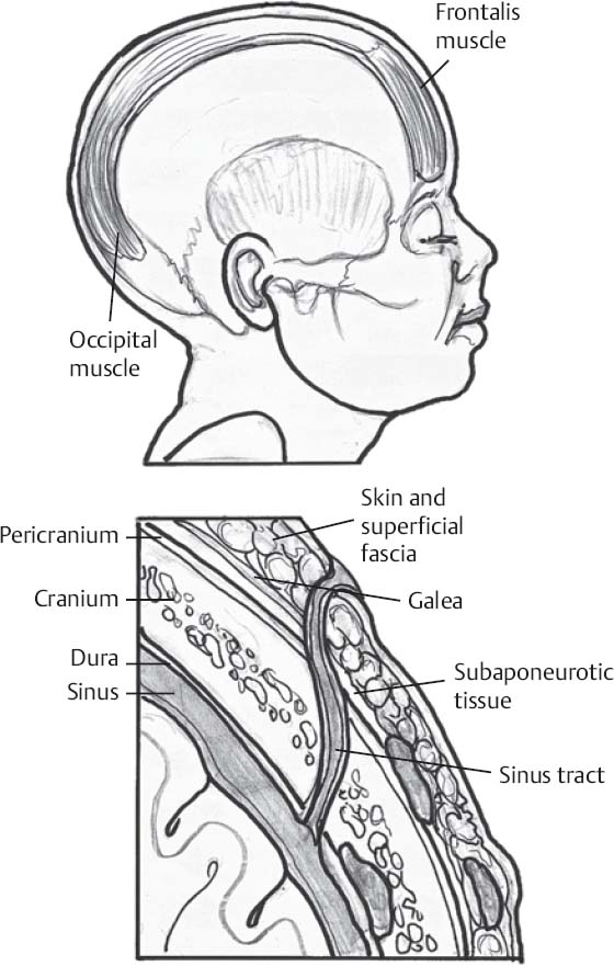
- Cranial sinus tracts:
- An epithelium-lined tract that extends from the skin surface at variable depths into tissues (Fig. 85.1)
- A persistent attachment that probably forms when cutaneous ectoderm does not separate completely from neuroectoderm during neural tube formation
- Found in either the midline nasal or occipital regions
- Associated with a variety of cutaneous findings: hair, hemangioma, dimple, or lipoma
- Occipital cysts are midline and extend subcutaneously in a caudal direction
- Those penetrating the calvarium enter the bone inferior to the torcula
- Dermoid or epidermoid cysts may be found anywhere along the sinus tract
- Often discovered following bouts of recurrent sterile (chemical) or bacterial meningitis
- Tracts should never be probed during physical examination to avoid introducing infectious materials intracranially
- Magnetic resonance imaging is best to visualize the sinus tract and level of extension, and to determine presence of intracranial abnormality such as a dermoid cyst
- Goal of surgery is elective total excision of the entire sinus and cyst (when present) prior to possible infectious complication
- An epithelium-lined tract that extends from the skin surface at variable depths into tissues (Fig. 85.1)
Equipment
- Major craniotomy tray
- Mayfield head holder
- High-speed drill
Operating Room Set-up
- Headlight
- Loupes
- Bovie cautery and bipolar cautery
< div class='tao-gold-member'>
Only gold members can continue reading. Log In or Register to continue
Related posts:


Stay updated, free articles. Join our Telegram channel

Full access? Get Clinical Tree


