Sensation
What You Find and What It Means
WHAT YOU FIND
Patterns of sensory loss
Sensory deficits (Fig. 22.1) can be classified into eight levels of the nervous system:
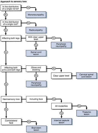
1. Single nerve: sensory loss within the distribution of a single nerve, most commonly median, ulnar, peroneal, lateral cutaneous nerve to the thigh. Distributions are illustrated in Chapter 21.
2. Root or roots: sensory deficit confined to a single root or a number of roots in close proximity—common roots in the arm C5, C6 and C7 and in the leg L4, L5 and S1. Distributions are illustrated in Chapter 21. When multiple nerve roots are involved in the lumbosacral spine (usually S1–S5 roots bilaterally), this results in a cauda equina syndrome with sensory loss in the perianal region and buttocks (saddle anaesthesiae) and the back of both thighs.
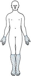
3. Peripheral nerve: distal glove and stocking deficit (Fig. 22.2).
4. Spinal cord: five patterns of loss can be recognised (Fig. 22.3):
• Complete transverse lesion: hyperaesthesia (increased appreciation of touch/pinprick) at the upper level, with loss of all modalities a few segments below the lesion (Fig. 22.3A).
• Hemisection of the cord (Brown–Séquard syndrome): loss of joint position sense and vibration sense on the same side as the lesion and pain and temperature on the opposite side a few levels below the lesion (Fig. 22.3B).
• Central cord: loss of pain and temperature sensation at the level of the lesion, where the spinothalamic fibres cross in the cord, with other modalities preserved (dissociated sensory loss)—seen in syringomyelia (Fig. 22.3C).
Related posts:


Stay updated, free articles. Join our Telegram channel

Full access? Get Clinical Tree


