Sinonasal Disease

Chapter 13 Sinonasal Disease


We follow the classic approach of reviewing the anatomy of the sinonasal cavity and then proceed to congenital, inflammatory, and neoplastic categories. There will be some redundancy as we describe clinical versus surgical considerations in some of these categories.


To appreciate the pathogenesis of sinusitis, you must understand the normal anatomic pathways of mucociliary clearance in the paranasal sinuses (Fig. 13-1). The cilia within the maxillary sinus propel the mucus stream in a starlike pattern from the floor of the maxillary sinus toward the ostium, situated superomedially. In approximately 30% of patients, a second accessory ostium to the maxillary sinus is present inferior to the major opening. From the maxillary sinus ostium, mucus from the maxillary antrum (the maxillary antrum and maxillary sinus are synonymous) gets swept superiorly through the infundibulum, which is located lateral to the uncinate process and medial to the inferomedial border of the orbit. The uncinate process, a sickle-shaped bony extension of the lateral nasal wall extending anterosuperiorly to posteroinferiorly, is rarely (<2.5% of patients) pneumatized itself. Occasionally, the uncinate process attaches to the lamina papyracea (the medial wall of the orbit). If it does so, the infundibulum does not have a superior opening, thus creating a blind pouch, the recessus terminalis. The hiatus semilunaris is a slitlike air-filled space anterior and inferior to the largest ethmoid air cell, the ethmoidal bulla, and right above the uncinate process. Mucus is passed through the hiatus semilunaris posteromedially via the middle meatus, a channel between the middle turbinate and the uncinate process, into the back of the nasal cavity to the nasopharynx, where it is subsequently swallowed.


The ostiomeatal complex (OMC) refers to the maxillary sinus ostium, the infundibulum, the uncinate process, the hiatus semilunaris, the ethmoid bulla, and the middle meatus—the common drainage pathways of the frontal, maxillary, and anterior ethmoid air cells.


The frontal sinuses drain inferomedially via the frontal recess (also called the frontoethmoidal recess). The frontal recess connotes the common drainage of the frontal sinus and the anterior ethmoid air cells. The frontal recess is the space between the inferomedial frontal sinus and the anterior part of the middle meatus. The frontal sinus and the anterior ethmoid air cells usually drain directly into the middle meatus via the frontal recess, or less commonly into the superior ethmoidal infundibulum, before passing to the middle meatus.


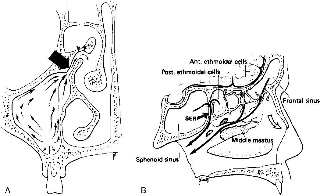
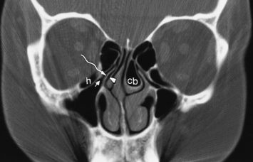
The most anterior ethmoid air cells located in front of the middle turbinate’s cribriform plate attachment are termed agger nasi cells. Agger nasi cells lie anterior, lateral, and inferior to the frontal recess. They are present in more than 90% of patients (Fig. 13-2). The roof of the ethmoid sinus is termed the fovea ethmoidalis.


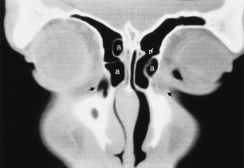
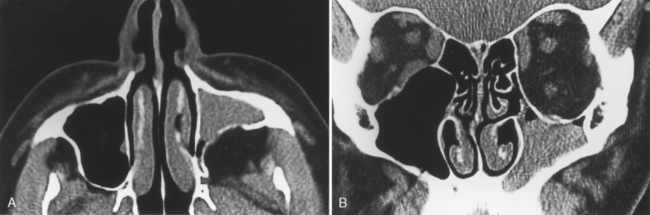
As stated earlier, ethmoidal bulla is the term used for the ethmoid air cell directly above and posterior to the infundibulum and hiatus semilunaris. A very large ethmoidal bulla can obstruct the infundibulum and hiatus semilunaris, leading to interference with the drainage of the maxillary and anterior ethmoid sinuses. When anterior ethmoid air cells are located inferolateral to the bulla, along the inferior margin of the orbit protruding into the maxillary sinus, they are termed Haller cells, maxilloethmoidal cells, or infraorbital cells. They are seen in 10% to 45% of patients. When greatly enlarged, Haller cells may narrow the infundibulum or maxillary sinus ostium (Fig. 13-3).


Between the ethmoidal bulla and the basal lamella (the lateral attachment of the middle turbinate to the lamina papyracea of the orbit) is the sinus lateralis. The sinus lateralis, comprising the suprabullar and retrobullar recesses, may open into the frontoethmoidal recess or into a space posterior to the bulla, the hiatus semilunaris posterioris.


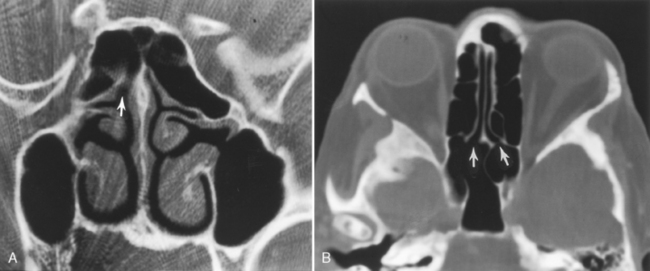
The posterior ethmoid air cells are located behind the basal (or ground) lamella of the middle turbinate and drain via the superior meatus, the supreme meatus, or other tiny ostia just under the superior turbinate. Ultimately, these ostia drain into the sphenoethmoidal recess of the nasal cavity (Fig. 13-4), from which the secretions pass to the nasopharynx. In some patients the most posterior ethmoid air cell may pneumatize into the sphenoid bone, superior to the sinus. This is termed an Onodi cell.


The nasal cavity typically has three sets of turbinates: the superior, middle, and inferior turbinates. Occasionally, one may identify a fourth superiormost turbinate, the supreme turbinate. An aerated middle turbinate, which usually communicates with the anterior ethmoid air cells, is termed a concha bullosa and is seen in approximately 34% to 53% of patients. Most people believe that, unless huge, the presence of a concha bullosa does not predispose to chronic sinusitis. Significant pneumatization of the inferior or superior turbinates is much less common (<10% of patients).


The nasal septum is the midline structure between the right and left turbinates. The nasal septum is composed of three parts: a cartilaginous anteroinferior portion; a bony posteroinferior portion known as the vomer; and a superoposterior bony portion, the perpendicular plate of the ethmoid bone. The nasal septum is aerated only rarely. Nasal septal deviation, however, is common, and bony spurs often develop at the apex of the deviation. Spurs may cause the sensation of nasal obstruction.


The nasolacrimal duct (see Chapter 10) courses downward from the lacrimal sac bordering the medial canthus, where it is in close association with agger nasi air cells. Inflammation of agger nasi cells may be associated with epiphora because of this close relationship. The duct subsequently runs in the anterior and inferior portion of the lateral nasal wall. Its ending opens below the inferior turbinate at the inferior meatus.


Paranasal Sinus Development


The frontal sinus is not present at birth, but pneumatization evolves from age 1 to 12 years. Growth over the orbital roof usually occurs from ages 4 to 8 years. Frontal sinusitis before age 4 years is therefore rare. On the other hand, the ethmoid sinuses are present at birth. Rapid expansion of the ethmoid air cells occurs during ages 0 to 4 years and again with the adolescent growth spurt from age 8 to 12 years. The ethmoid sinus is usually the source of infection in childhood sinusitis. This may also lead to orbital cellulitis or subperiosteal abscesses in kids.


The maxillary antrum is also present though small at birth. Its growth continues to age 14 years, but it can be influenced by dental development. Tooth buds will be seen between the maxilla and the aerated sinus and obviously can prohibit an anterior antrostomy in these youngsters.


The sphenoid sinus begins its pneumatization at about age 2 years, and the growth is slower and more delayed than that of the other sinuses. The ultimate size of the sphenoid sinus is quite variable. Remember the development of the sinuses by this mnemonic: Embattled military fought Saddam (ethmoid, maxillary, frontal, sphenoid).


Note that the OMC is developed at birth, and functional endoscopic sinus surgery (FESS) is an option for chronic childhood sufferers of sinusitis. The mucosa in the infant is somewhat redundant and easily congested. Therefore, mucosal thickening should not be assumed to be due to sinusitis in a crying child. The clinical evaluation is paramount here. Up to 60% of asymptomatic infants can have complete or near-complete opacification of their sinuses.


IMAGING MODALITIES


Sinonasal imaging has progressed methodically as each new generation of imaging modality has encroached on the domain of the former generation. Although plain films once served as the most commonly ordered study to evaluate the sinonasal cavity, computed tomography (CT) has now supplanted plain films, because the endoscopic sinus surgeon has required greater anatomic precision. During the 1970s and 1980s, FESS replaced the more traditional Caldwell-Luc and maxillary antrostomy procedures for treating chronic sinusitis.


The plain-film examination, which consists of a Waters (brow-up anteroposterior [AP] view), Caldwell (frontal AP view), lateral, and submental vertex view, is as anachronistic in sinonasal imaging as a stethoscope is for evaluation of coronary artery disease. The drawback of overlapping structures makes the evaluation of the OMC, anterior ethmoid sinus, middle meatus, and sphenoid sinus grossly limited with plain films.


Functional endoscopic sinus surgery is done via an intranasal endoscope rather than with an external approach, so surgeons must know where they are at all times to prevent complications such as orbital or intracranial entry, particularly when they are operating posteriorly in the sinonasal cavity. CT serves as the road map for this procedure. The goal of FESS is to maintain the normal mucosa of the sinonasal cavity and to preserve the natural pathway of mucociliary clearance. FESS does not attempt to strip the mucosa clean as was sometimes performed in a Caldwell-Luc procedure, so mucociliary motility is preserved. Also, rather than creating an alternative egress of mucus from the maxillary sinus, as in an inferior meatal antrostomy (the Caldwell-Luc procedure), FESS enlarges the natural ostia and passageways of the paranasal sinuses. Whereas in the past maxillary and frontal sinusitis were thought to be the primary processes in patients with chronic sinu-sitis, it is now believed that these sinuses are secondarily obstructed because of disease in the OMC. The classic theory of FESS is that disease at the ostium and inferior infundibulum obstructs the maxillary sinus, whereas disease in the middle meatus and posterior infundibulum obstructs the frontal and anterior ethmoid air cells. Therefore, surgery is directed toward removing potential obstacles to mucociliary clearance at the OMC. Persistence of chronic sinusitis after nasoantral windows is usually due to anterior ethmoid disease. Therefore, amputation of the uncinate process, enlargement of the infundibulum and maxillary sinus ostia, and creation of a common unobstructed channel for the anterior ethmoid air cells are common practices in FESS. FESS may also include complete or partial ethmoidectomies with recurrent disease.


For the FESS surgeon, coronal CT is ideal because it simulates the appearance of the sinonasal cavity from the perspective of the endoscope. At present coronal reconstructions of axial CT data from multidetector spiral CT scans are nearly as good as direct coronal images and may be able to eliminate some dental amalgam artifact that otherwise is present. To eliminate the effects of reversible sinus congestion, patients undergoing CT for evaluation of chronic sinus disease are best scanned 4 to 6 weeks after medical therapy and not during an acute infection. Some radiology departments also administer nasal spray decongestants or antihistamines to reduce reversible mucosal edema before the patients are placed in the scanner. Even with this preparation, surgeons claim that in approximately 10% of cases with “normal CT scans” they find endoscopic evidence of significant sinusitis.


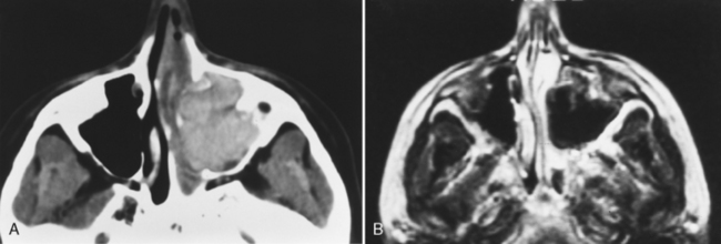
Magnetic resonance (MR) examination of the sinonasal cavity can be performed in a standard head coil or, for more precise anatomic resolution, with a surface coil placed over the anterior part of the face. Both T1-weighted images (T1WI) and T2-weighted images (T2WI) are required because of the variability of signal intensity of sinonasal secretions caused by protein concentration. Fat-suppressed enhanced T1WI is employed for the evaluation of complicated sinusitis or for suspected neoplastic disease. Differentiating tumors from infections of the sinonasal cavity may be best achieved with enhanced MR: Infected mucosa enhances in a peripheral fashion, whereas tumors usually enhance solidly and centrally (Fig. 13-5).


CONGENITAL DISORDERS


Choanal Atresia


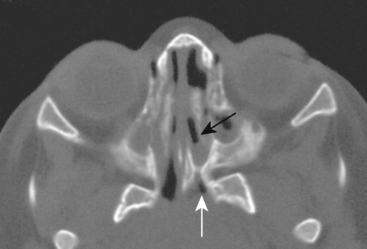
Choanal atresia is usually diagnosed in infancy because neonates are obligate nose breathers as they suck on a bottle or breast (Box 13-1). The child is seen with respiratory distress. Although the diagnosis can be suggested by the inability to pass a nasogastric tube through the nose, imaging is necessary to determine whether the obstruction is membranous (15% of cases) or bony (85%) and whether other congenital central nervous system (CNS) or non-CNS anomalies are associated (50%). In addition to the narrowed posterior choana, look for thickening of the vomer (Fig. 13-6). The posterior choanal opening should be more than 0.5 cm wide in neonates, 1 cm in adolescents. The vomer should measure less than 0.34 cm in children under age 8 years. Rather than atresia, some patients have mere stenosis of the passageway. Often, unilateral choanal atresia may escape detection into adulthood. Patients may be unaware of the hyposmia often associated with this disorder.


A dacryocystocele or piriform aperture stenosis may mimic choanal atresia clinically.


Dermoids, Sinus Tracts, and “Gliomas”


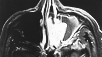
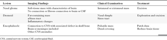
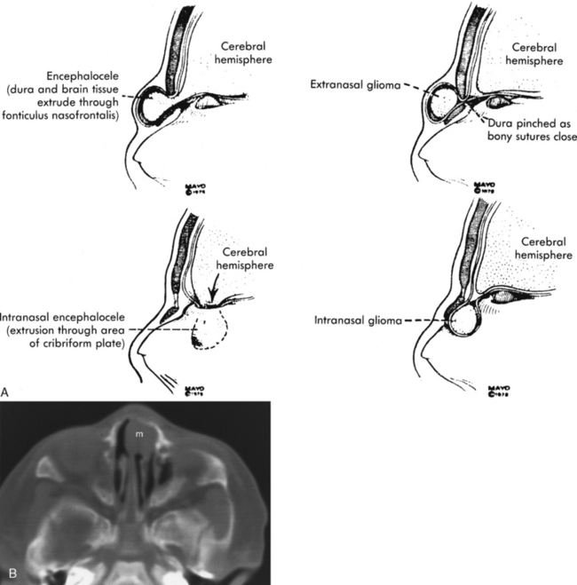
Congenital lesions of the sinonasal cavity include congenital encephaloceles, dermoid cysts, sinus tracts, and nasal gliomas (Table 13-1). These lesions occur as an abnormality in the process of invagination of the neural plate. In embryogenesis, the dura of the brain contacts the dermis at the nasion region as the neural plate retracts. Normally, the dermal connection regresses; when it does not, one of these lesions may develop. A cerebrospinal fluid (CSF) connection to the intracranial contents is maintained with meningoencephaloceles (see Chapter 9), whereas the connection is fibrous only with a nasal glioma (Fig. 13-7). Nasal gliomas are NOT neoplasms but congenital anomalies. They are extranasal more commonly than intranasal. (What an oxymoron—nasal gliomas are usually extranasal and are not gliomas.) Most patients with dermoid sinus tracts have a pit in the middle of the nose. Dermoid cysts occur more commonly than tracts; however, tracts may cause more severe symptoms because of their intracranial connection in 25% of cases. Thus meningitis, osteomyelitis, and intracranial abscesses may occur in the setting of dermoid tracts.


Hypoplastic Maxillary Antrum


Congenital hypoplasia of the maxillary sinus occurs in 9% of patients. On plain films, a hypoplastic maxillary antrum (appearing denser) can simulate sinus opacification, but the CT will not fall into this trap.


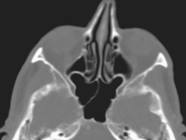
Bony changes that suggest the diagnosis of a hypoplastic antrum are listed in Box 13-2. Causes of overexpansion of paranasal sinuses are listed in Box 13-3. In the differential diagnosis of sinus hypoplasia is the “silent sinus syndrome,” or maxillary sinus atelectasis. In this entity, ostial obstruction from chronic sinusitis leads to chronic negative pressure, which leads to hypoventilation, which, over time, reduces the sinus’ volume, hence sinus atelectasis. Patients present with enophthalmos (not sinus symptoms, strangely enough) as the orbital floor becomes depressed, the maxillary walls retract centripetally, and the retromaxillary fat fills the space left by the atelectatic sinus. The CT shows the retracted maxillary sinus walls in association with a small-volume, opacified sinus (Fig. 13-8).


INFLAMMATORY LESIONS


Sinusitis


From the standpoint of a public health hazard, sinusitis ranks as one of Americans’ most common afflictions. It is estimated that more than 31 million people in the United States are affected by sinus inflammatory disease each year and that 16 million visits to primary care physicians annually are for sinusitis and its complications. Adults average two to three colds per year, and 0.5% of viral upper respiratory infections are complicated by sinusitis. Overall, Americans spend more than $150 million per year for over-the-counter cold and sinusitis medicines, $100 million of which is for antihistamine medications.


Most cases of acute sinusitis are related to an antecedent viral upper respiratory tract infection. With mucosal congestion as a result of the viral infection, apposition of mucosal surfaces results in obstruction of the normal flow of mucus, which results in retention of secretions, creating a favorable environment for bacterial superinfection. The ethmoid sinuses are most commonly involved in sinusitis, possibly because of their position in the “line of fire” as inspired particles collide with and irritate the fragile ethmoid sinus lining. The bacterial pathogens responsible for acute sinusitis include Streptococcus pneumoniae, Haemophilus influenzae, β-hemolytic streptococci, and Moraxella catarrhalis. In the chronic phase Staphylococcus, Streptococcus, corynebacteria, Bacteroides, fusobacteria, and other anaerobes may be responsible. The fungi that may infect the sinuses include Aspergillus, Mucor, Bipolaris, Drechslera, Curvularia, and Candida.


Anatomic Considerations


Several issues must be addressed when a patient’s CT is evaluated before FESS. Is the uncinate process apposed to the medial orbital wall (an atelectatic infundibulum)? If so, its vigorous removal may result in orbital penetration. Are there areas of dehiscence in the lamina papyracea, or do the orbital contents protrude into the ethmoid sinus (both of which may lead to unintentional orbital entry from the ethmoid sinus)? Defects in the lamina papyracea have been reported in 5% to 10% of autopsy specimens. Because orbital hematomas are the most common orbital complication of FESS, it is important to identify any gaps in the lamina papyracea. CT obviously is the best means for identifying the thin medial bony wall of the orbit.


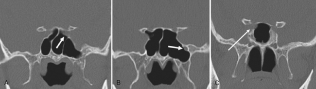
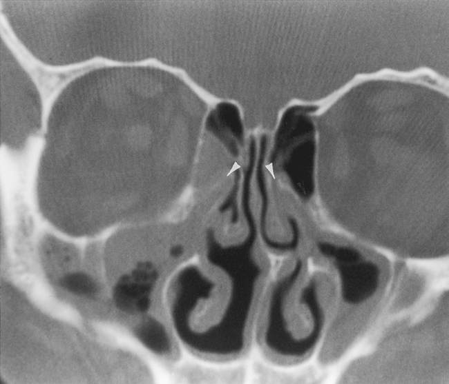
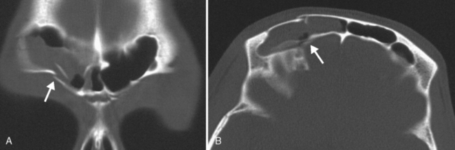
Are there areas of dehiscence in the cribriform plate and sphenoid sinus walls? Remember that there is an attachment of the middle turbinate to the cribriform plate. If the surgeon tugs too hard, the cribriform will fall and down will come CSF or brain through the new foramen in the skull base. The potential for intraorbital, intracranial, carotid, or optic nerve perforation at the time of surgery depends on these anatomic variants, found in 4% to 15% of patients. Three percent of people have optic nerves that are in contact with the posterior ethmoid wall—most course along (90%) or through (6%) the sphenoid sinus. An area of bony dehiscence seen on CT is present in 24% of optic nerves, putting it at risk (Fig. 13-9). There have been limited reports of optic nerve transection during sphenoethmoidectomy from an intranasal approach, and dehiscence of the sphenoid wall may be a predisposing factor. An intersinus septum in the sphenoid sinus that attaches to the carotid canal is important to recognize preoperatively and is typically best identified in the axial view (Fig. 13-10). Overvigorous removal of such an intersinus septum during surgery may result in carotid laceration.


Appearance on Imaging


In addition to commenting on the normal anatomic variations in the CT report, the radiologist should identify areas of mucosal thickening and sinus passageway opacification. It has come to be accepted that the location of sinusitis is more important in producing a patient’s symptoms than the extent of the sinusitis. Therefore, a subtle area of opacification in the infundibulum of the OMC may cause more pain and discomfort than nearly complete opacification of the maxillary sinus with a mucus retention cyst or polyp.


OMC opacification correlates well with the development of sinusitis (Fig. 13-11). The positive predictive value of infundibular opacification for the presence of maxillary sinus inflammatory disease is approximately 80%. When the middle meatus is opacified, the maxillary and ethmoid sinuses show inflammatory change in 84% and 82% of patients, respectively. The specificity of middle meatus opacification for maxillary or ethmoid sinus disease is more than 90%. These findings support the contention that obstruction of the narrow drainage pathways leads to subsequent sinus inflammation.


Some head and neck radiologists categorize recurrent inflammatory sinonasal disease into five patterns: (1) infundibular, (2) ostiomeatal unit, (3) sphenoethmoidal recess, (4) sinonasal polyposis, and (5) sporadic or unclassifiable disease. The infundibular pattern is seen in 26% of patients and refers to isolated obstruction of the inferior infundibulum, just above the maxillary sinus ostium. Limited maxillary sinus disease often coexists with this pattern, whereas the ostiomeatal unit pattern, seen in 25% of cases, often has concomitant frontal and ethmoidal disease. The ostiomeatal unit pattern is designated when middle meatus opacification is present. Sphenoethmoidal recess obstruction occurs in 6% of cases and leads to sphenoid or posterior ethmoid sinus inflammation. When the sinonasal polyposis pattern is present, enlargement of the ostia, thinning of adjacent bone, and opacified sinuses are usually seen.


As far as the degree of sinus disease, some use the Lund Mackay staging system, in which the frontal, anterior ethmoid, posterior ethmoid, sphenoid, and maxillary sinus, as well as the OMC on each side, are each assigned a grade. The grades are 0 for clear, 1 for partial opacification, and 2 for complete opacification.


The presence of air-fluid levels is more typically associated with acute sinusitis than with chronic inflammatory disease. In cases of suspected acute sinusitis, air-fluid levels or complete opacification of a sinus is present in 63% of cases (Fig. 13-12). Using these two criteria (air-fluid levels and complete opacification), CT has a 90% positive predictive value for acute sinusitis. Bubbles of air mixed with soft tissue in the sinus are another predictor of acuity. Of course, acute sinusitis may be superimposed on chronic changes. The findings suggestive of chronic sinusitis include mucosal thickening, bony remodeling, polyposis, mucus retention cysts, and bone thickening secondary to osteitis from adjacent chronic mucosal inflammation (Fig. 13-13). Some people exclude mucosal thickness of less than 3 mm as normal passive mucosal congestion that does not indicate inflammation, particularly in children.
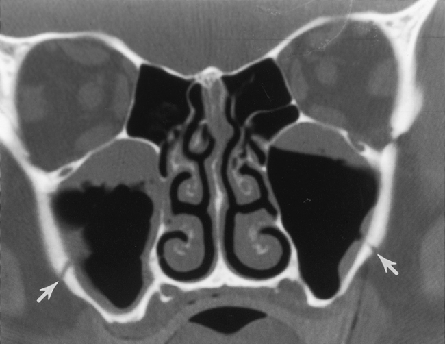
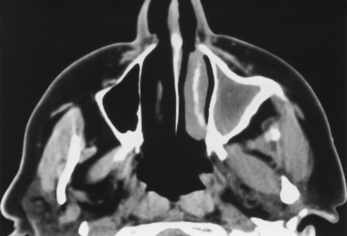


Hyperdense secretions on CT may be due to six main causes: (1) inspissated secretions, (2) fungal sinusitis, (3) hemorrhage in the sinus, (4) polyps, (5) mucocele, and (6) calcification. The hyperdense sinus may be the only clue to fungal sinusitis and is an important feature to note. However, chronic sinusitis infected with bacteria occasionally is hyperdense on CT, particularly in patients who have very long-standing disease or cystic fibrosis. The hyperdense sinus often corresponds to the hypointense sinus on T2WI (Fig. 13-14). Nonetheless, say “a fungus is among us” if you see dense secretions.


One may also see calcifications in the maxillary antra that may indicate fungal sinusitis; however, once again, the finding may also be present with nonfungal inspissated secretions (though not to the same extent). Here are a few tips: If the intrasinus calcification is central and is fine, punctuate-like, it is most likely due to fungi. If the calcification is peripheral, curvilinear, and eggshell-like, it is probably nonfungal. If you measure the Hounsfield units (HU) you can gain some specificity to the “hyperdense sinus.” Those with HUs greater than 2,000 have a 93.3% chance of maxillary sinus aspergillosis. The mean CT density of the sinus concretions without aspergillosis is 778 HU.


The following features of the “calcified” sinus mass have been reported. A single discrete hyperdensity is most likely to be an inflammatory mass (aspergilloma, rhinolith), but multiple discrete calcifications could be seen in tumors (enchondromas, inverted papillomas, meningiomas) or inflammatory lesions. If the process is diffusely hyperdense with a well-defined margin, think of a fibro-osseous lesion, but if it is poorly marginated, consider a high-grade sarcoma (chondrosarcoma, osteosarcoma). Although calcification is not unusual in inverted papillomas it is more likely to be residual bone, not calcifications. On the other hand, esthesioneuroblastomas have intrinsic calcifications.


A rhinolith (stone in the nose) is often due to a foreign body that has become lodged in the nose and has slowly calcified. This may occur in the setting of traumatic or recreational nasal septum injuries.


While you are evaluating coronal CT scans for sinusitis, do not forget to look at four other related areas: the teeth, the sella, the nasopharynx, and the temporomandibular joints. Pain from odontogenic infections or carious teeth may be mistakenly attributed to sinusitis. Therefore, check the maxillary teeth for areas of demineralization or periapical cysts as you check for sinusitis. By the same token, of those patients referred for the teeth, mandible, and maxilla with periodontal disease, 60% had maxillary sinusitis, as opposed to those without periodontal disease, who had just a 25% rate of maxillary disease. It is very useful to observe the displacement of the maxillary sinus wall to distinguish odontogenic cystic lesions, which displace the inferior antral wall superiorly, from maxillary sinus lesions such as mucoceles, which push the bone down.


Malocclusions or temporomandibular joint degenerative change may indicate a maxillofacial pain syndrome that may simulate pain from sinusitis. Look for narrowing or sclerosis around the joint. We are also surprised at the high rate of incidental sellar lesions and nasopharyngeal carcinomas shown by plaintiff lawyers that are missed by the unwary radiologist reading sinus CT scans.


Screening sinus CTs are often performed before bone marrow transplantation (BMT) in patients with hematologic malignancies. Of those showing severe sinusitis on initial screening CT scans, two thirds experience clinical sinusitis after BMT. Forty percent of patients with moderate sinus abnormalities on pre-BMT CT scans develop clinical sinusitis during their post-BMT course, compared to 23% with normal pre-BMT CT scans. The presence of sinusitis on pre-BMT studies is also associated with a trend toward overall decreased survival. Premedicate for sinusitis before BMT is given.


Trauma often affects the walls of the paranasal sinuses. Remember that the floor of the orbit serves as the roof of the maxillary sinus, so fractures there cause blood levels in the sinuses. Similarly, the medial wall blowout fracture affects the ethmoid sinus, and orbital roof fractures may affect supraorbital ethmoid cells or the frontal sinus. Direct blows to the forehead may cause inward displacement fractures of the frontal sinuses (Fig. 13-15). Skull base fractures may cross the sphenoid sinus. These air-filled spaces are not necessarily the best buttresses against trauma, being very thin and with nothing but air between them and the directed force.


< div class='tao-gold-member'>

Stay updated, free articles. Join our Telegram channel

Jul 22, 2016 | Posted by in NEUROLOGY | Comments Off on Sinonasal Disease

Full access? Get Clinical Tree

Get Clinical Tree app for offline access