Midazolam
Propofol
Benzodiazepine derivative
2-6 Disopropylphenol
Large therapeutic range
Small therapeutic range
Active etabolites (accumulation)
No accumulation
Functional half life 45 min
Functional half life 4–6 min
Elimination half life 150 min
Elimination half life 55 min
Central muscle relaxation
Central breathing depression
Paradoxal reaction in 1 %
Hypopharyngeal reflex depression
Subjects with an AHI below 30, or to be more accurate patients with a supine AHI below 30 and with good health (ASA I or II), can undergo midazolam-induced sleep endoscopy in the clinic. Midazolam is administered by the ear, nose, and throat surgeon (the presence of an anesthetist is not obligatory) or by an anesthetist. Sleep is induced by giving midazolam intravenously, slowly titrated up to 0.07 mg/kg per patient, followed by a saline flush. If insufficient, a bolus of 1–2.5 mg is given (a maximum of 7.5 mg per patient). Patients who are extremely nervous or who habitually use antidepressants or sedatives may need an extra bolus.
Previous studies reported that propofol did not change the respiratory pattern nor significantly influence the AHI, but did interfere with the sleep architecture, specifically, reduction in rapid eye movement (REM) sleep in patients undergoing propofol-induced sleep endoscopy [9]. Respiratory and sleep parameters did not change significantly during diazepam-induced sleep endoscopy in comparison with natural sleep either, except for a small increase in the apnea index and a minor change in the duration of the longest apnea and REM sleep [10].
Anesthetic depth is of key importance. The ideal concentration of the respective sedative is variable according to an individual’s susceptibility to the sedative effect of the drug. Slow stepwise induction is key to avoid oversedation, resulting in a loss of consciousness. The latter is related to a decrease of upper airway muscle tone and an increase in pharyngeal critical closing pressure.
Once the patient has reached a satisfactory level of sedation, a flexible endoscope (e.g., 3.5 mm) lubricated and coated with anticondense is introduced into the nasal cavity.
The nasal passage, nasopharynx, velum, tongue base, epiglottis, and larynx are observed. The levels of snoring and/or obstruction are assessed.
During the DISE, maneuvers such as a chin lift (a manual closure of the mouth) or a jaw thrust (or Esmarch maneuver) should be performed, with reassessment of the airway after each maneuver. A jaw thrust is a gentle advancement of the mandible by up to 5 mm, mimicking the effect of a mandibular repositioning device. It is thought that, using DISE, one can predict the likelihood that an appliance would be effective by examining the changes in the airway. Although the effects during sedation may not be identical to those of natural sleep, the distance of protrusion can be measured and can inform decisions about the necessary degree of mandibular repositioning with an appliance.
In patients with an insufficient effect of a mandibular repositioning device, DISE can be performed without the device both in and out, to assess obstruction site(s) and surgical alternatives.
VOTE Classification
There is a wide range of systems which describe the complex interactions of upper airway structures during DISE. Some exclude the epiglottis; others try to group multiple structures together in various combinations. However, there is no universally used DISE scoring system—hence one is needed.
We therefore recently proposed the VOTE classification system for reporting DISE findings, with a focus on the primary structures that contribute to upper airway obstruction, either alone or in combination: the velum, oropharyngeal lateral walls (including the tonsils), tongue, and epiglottis [11].
The VOTE classification may be an oversimplification that overlooks some interactions, but we believe it is a foundation for further study of pharyngeal obstruction in OSA and for assessment of the response of upper airway structures to directed interventions. Our experience suggests that a focus on structures could help answer two central questions: treatment selection and the association between DISE findings and treatment outcomes—for surgery, mandibular repositioning devices, or combined therapy. The VOTE classification represents a common language to describe the patterns of obstruction during DISE and may ultimately determine treatment interventions (Fig. 1).
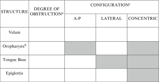
Fig. 1
The VOTE classification. For each structure, there should be a classification as to the degree of obstruction and configuration of obstruction. Open boxes reflect the potential configuration that can be visualized related to a specific structure. Shaded boxes reflect the fact that a specific structure–configuration cannot be seen (e.g., oropharynx lateral walls in an anteroposterior direction). A-P anteroposterior. aDegree of obstruction has one number for each structure: 0, no obstruction (no vibration); 1, partial obstruction (vibration); 2, complete obstruction (collapse); X, not visualized. bOropharynx obstruction can be distinguished as related solely to the tonsils or including the lateral walls, with or without a tonsillar component. cConfiguration noted for structures with degree of obstruction greater than 0
The most common and well-known sites of obstruction and vibration are located in the soft palate and the lateral pharyngeal walls, including tonsils and the base of tongue. Obstruction at epiglottic level occurs less often but has clinical significance. Previous large series of DISE in patients with OSA reported a majority of multilevel obstruction, a retropalatinal as well as retrolingual obstruction in a large percentage of cases. In general, a unilevel obstruction is more common in patients with mild OSA, while in severe OSA, a multilevel obstruction is more likely, being the “culprit” for the severity of the OSA.
The subsequent surgical treatment with different, site-specific procedures will not be discussed here. For many years, surgical evaluation techniques have focused on categorizing patients first according to the Fujita classification system that encompasses the two primary regions of pharyngeal upper airway obstruction: the palatal/velopharyngeal and hypopharyngeal/retrolingual regions. However, there are two major limitations of a region-based classification. First, there is substantial anatomical overlap between these regions, including the extension of the lateral pharyngeal walls throughout the length of the pharynx and the physical overlap of the tongue and soft palate. Second, a region-based approach may not determine surgical treatment adequately. For example, in patients with hypopharyngeal/retrolingual obstruction, the oropharyngeal lateral walls, tongue, and epiglottis can each play a more prominent role.
One of the biggest advantages of DISE is the individual analysis, which allows patient-specific and site-specific therapies according to location and degree of obstruction. Maurer et al. have found that in difficult cases, the therapeutic plans are changed after DISE in up to 75 %. Although we have the impression that surgical success rates in patients selected by DISE are better than average, this has to be confirmed in more studies.
The Structures of the VOTE Acronym
Our experience with over 7,500 DISE examinations suggests that a selected group of structures contribute to upper airway narrowing and/or obstruction in sleep-disordered breathing, individually or in combination. The VOTE classification (Fig. 1) evaluates these structures and the degree of airway narrowing.
Velum
Velopharyngeal obstruction occurs at the level of the soft palate, uvula, or lateral pharyngeal wall tissue at the level of the velopharynx. Because these three structures are not entirely distinct entities—both anatomically and on DISE—we have grouped them together. Airway closure related to the velum can occur with collapse in an anteroposterior or concentric configuration, but rarely in a lateral configuration.
Oropharyngeal Lateral Walls Including the Tonsils
The oropharyngeal lateral walls include two structures: the tonsils and the lateral pharyngeal wall tissues that include musculature and the adjacent parapharyngeal fat pads. Both structures collapse in a lateral configuration, although this may occur in combination with collapse of other structures, with a resulting concentric pattern. In the presence of lateral wall collapse, it can be difficult (but certainly not impossible) to determine whether the tonsils or lateral walls are playing a significant role, reflecting potential subtypes; importantly, the distinction can have important implications for treatment selection and outcomes. While the VOTE classification is largely based on DISE findings alone, the examination of tonsil size and lateral pharyngeal wall tissues during routine oral cavity examination are also important. Obstruction related to the oropharynx can only occur with collapse in a lateral configuration.
Tongue Base
Tongue base obstruction is a common DISE finding, and it results in anteroposterior narrowing of the upper airway. In natural sleep, there is a reduction in muscle tone of the tongue, especially during non-REM and REM sleep that is more pronounced in OSA patients compared to healthy individuals. Airway closure related to the base of tongue occurs with collapse in an anteroposterior direction.
Epiglottis
Epiglottic collapse occurs in one of two configurations, anteroposterior or lateral, but not concentric. Anteroposterior collapse can result with folding of the epiglottis with what appears to be decreased structural rigidity of the epiglottis or with an apparent posterior displacement of the entire epiglottis against the posterior pharyngeal wall, with normal epiglottic structural integrity. The second pattern, a lateral folding or involution, is consistent with a central vertically oriented crease of decreased rigidity of the epiglottis. The epiglottis may be under-recognized as a factor in patients with sleep-disordered breathing, and a substantial proportion of patients with OSA do demonstrate a significant epiglottic contribution to airway obstruction during DISE. DISE may provide a unique assessment of the epiglottis, as its apparent role has not been demonstrated as clearly demonstrated with other evaluation techniques (Fuyita, Mallampati/Friedman).
Related posts:
 of Positional Therapy: Transition from Tennis Balls to New Devices
Therapy and Palatal Surgery
and Future Perspectives
of Positional Therapy: Transition from Tennis Balls to New Devices
Therapy and Palatal Surgery
and Future Perspectives
 in Site of Obstruction in Obstructive Sleep Apnea Patients According to Sleep Position
 of Upper Airway Surgery on Positional Change During Sleep
 Impact of Body Weight Changes on Body Posture Dominance in Adult Obstructive Sleep Apnea Patients
Stay updated, free articles. Join our Telegram channel

Full access? Get Clinical Tree


