Rupture of the aorta or its branches is associated with reduced elasticity of the aorta wall in older patients and with the sharp lordotic angle and elongation of the anterior column. Generally, accepted theories include stretch of calcific nondistensible and tethered vessels creating internal and media tears leading to rupture and aneurysms. Although this risk is small [6–8], many surgeons choose pedicle subtraction osteotomy (PSO) to avoid this complication. Vascular injury has been reported if the opening wedge was performed at L1-L2 or L2–L3 [6, 8, 9]. Reviewing these reports and postmortem findings reveals 2 cases of rheumatoid spondylitis treated before surgery with radiograph therapy, in which adherence between the aorta and the underlying anterior longitudinal ligament resulted in a complete transverse tear in the posterior wall of the aorta after manual osteoclasis and nonsurgical or surgical correction [8, 9]. Such preoperative therapy was not conducted presently. We did not find any reference in the literature that the presently observed etiologies are linked to aortic-longitudinal ligament adherence. In the other 2 cases involving ankylosing spondylitis kyphotic patients with atheromatous calcification of their abdominal aorta [6], the vascular injuries involved SPO-related aortic wall tear and dissection of media. However, based on our observations of the absence of aortic injury in 354 patients treated with SPO including 101 patients with atheromatous calcification of abdominal aorta, apical lordosation osteotomy [11], or COWO [12] for correction of sagittal imbalance or kyphosis via an anterior open wedge and lengthening of the anterior column, the aorta can likely tolerate stretch and lengthening very well, even when complicated by atheromatous calcification [13].
Many series used SPO in which the spine is osteotomized through the posterior elements and corrected by direct pressure on the osteotomy site. The upper body and legs are extended to form a hollow cavity between the patient’s ventral trunk and the surgical table, and the pressure causes the ossified anterior vertebral column to fracture. This often occurs with a sudden snap, which suddenly stretches the aorta opposite the anterior opening wedge and might injure the aorta. The osteoclasis created by this pressure might avulse a bone fragment from the vertebral body to form a spike, and the aorta may be tensed most at the spike while the anterior wedge is opened, which might lead to aortic damage. In our SPO patients, the osteoclasis usually occurred at the intervertebral disc at the level of osteotomy during performed posterior osteotomy by gravity on the patient’s trunk or by light pressure on the osteotomy site after osteotomy. If the ossified anterior vertebral column was too hard to be fractured by light pressure, a fluoroscopically guided blunt ended osteotome was placed through the intervertebral disc at the level of the osteotomy. Then, osteoclasis was formed by gentle manipulation. The osteoclasis thus formed occurs at anterior disc space. Correction should not be started without assured osteoclasis and is accomplished by a slow and finely controlled closure of the osteotomy. As the posterior wedge is closed, correction occurs in the anterior vertebral column by opening of the anterior disc space with smooth edges (Fig. 7.2). All these managements for SPO were to minimize the risks of vascular complications and seemed effective because none of our SPO patients had a vascular injury [14]. This maneuver also results in an anterior column defect occasionally requiring reconstruction with anterior column support. In some cases, if substantial correction is achieved with an SPO, it may be necessary to graft the disc space anteriorly. In most cases, if a moderate correction is achieved at each level and the posterior column is closed bone-on-bone centrally and laterally, the SPO is likely to heal and anterior grafting and reconstruction is not needed. The technique is executed by resecting a “V”-shaped portion of the posterior elements (lamina and spinous process) and fusion mass between adjacent pedicles and correction is achieved by posterior column closing wedge and anterior column opening wedge. The effectiveness of this technique is much improved when it is applied to those patients who do not have an anterior column fusion (Fig. 7.3).
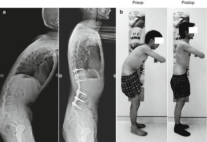
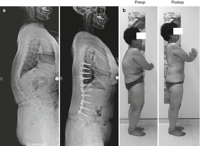

Fig. 7.2
(a) Preoperative lateral radiograph shows a 31-year-old male with ankylosing spondylitis related flexion deformity. Postoperative lateral radiograph shows correction of the deformity with SPO at L2–L3. The edge of the opening wedge is smooth. (b) Preoperative and postoperative clinical appearance

Fig. 7.3
(a) Lateral view of a 43-year-old female suffering a degenerative thoracolumbar kyphosis. Postoperative lateral radiograph shows correction of the deformity with SPO at L1–L2 where do not have an anterior column fusion. Posteriorly weakening the disc and anterior longitudinal ligament with a blunt-ended osteotome can facilitate osteoclasis. (b) Preoperative and postoperative clinical appearance
7.1.2 Ponte Osteotomy (PO)
The Ponte osteotomy (PO), described by Alberto Ponte in 1987 [15, 16], was described as a multilevel thoracic procedure to treat flexible thoracic kyphosis (Fig. 7.4). It is executed by undertaking an aggressive resection of the unfused facet joints, lamina, intraspinous ligaments, and ligamentum flavum at each level. Since the “osteotomy” is applied to the unfused spine, it could be argued that it is not an osteotomy in the true sense of the term. However, since it does require significant and specific osseous resection to be effective, the fused versus unfused status of the spinal segment is probably irrelevant. The PO resection can be narrow, amounting to an aggressive facetectomy or as radical as a complete resection of the posterior elements from pedicle to pedicle at each level. Since the technique is performed on the unfused spine, there is a mobile disc at each level, which may at times be difficult to discern. If there is no bony bridge anteriorly, then it is feasible. If there is a fine bony bridge, it can potentially be broken with closed osteoclasis and a PO turns into a SPO. Another difference between the SPO and the PO is that it is performed at multiple levels. This spreads the angular correction and anterior column translation, if any, over multiple levels. When using this technique, it results in an overall “closing wedge” effect. The anterior column remains supported by its undisturbed physiologic structures (discs and ligaments), leaving a stable spine after posterior column reconstruction. Additional anterior column procedures are unnecessary. This differs from the SPO’s anterior column “opening wedge” and posterior column “closing wedge” effect. The middle column functioning as a pivot point during correction of sagittal imbalance or kyphotic deformity with SPO or PO results in the least change of length and risk of injury of neural tissue comparing with PSO and vertebral column resection (VCR).
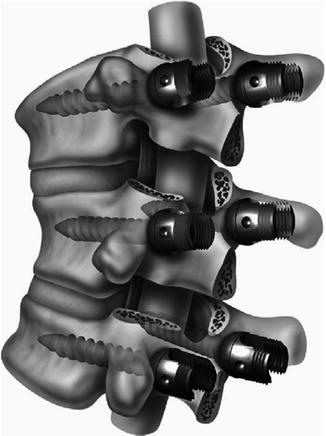

Fig. 7.4
Ponte osteotomy (Reproduced with the permission of Harry L. Shuftlebarger, MD)
Multiple osteotomies will be required to achieve significant correction since a PO may only provide a few degrees of correction per level (Fig. 7.5). In addition, the technique requires a mobile anterior and middle column to be useful. It may at times be difficult to discern. If there is no bony bridge anteriorly, then it is feasible. If there is a fine bony bridge, it can potentially be broken with closed osteoclasis. If there is a thick, solid bony bridge, it will not budge without release. While working best on long flexible kyphosis, even relatively stiff curves may be effectively treated with this technique. The PO may also be useful when performed above and below more aggressive osteotomies to provide a harmonious transition between areas of maximum and minimum kyphosis. PO can also be added as an afterthought to add a few more degrees of correction above or below a major deformity correction (Fig. 7.6).
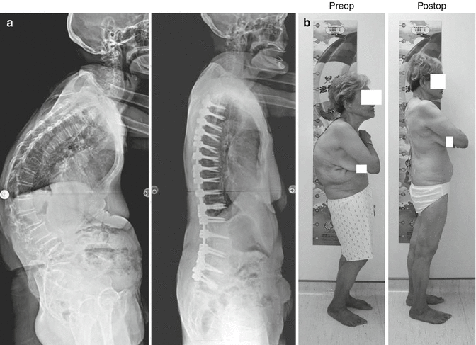
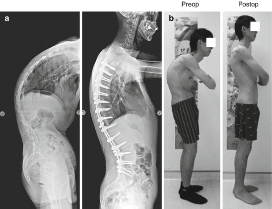

Fig. 7.5
(a) Lateral view of a 70-year-old female suffering senile kyphosis. Postoperative lateral radiograph of spine showing correction of deformity with multiple POs. (b) Preoperative and postoperative clinical appearance

Fig. 7.6
(a) Lateral radiograph of a 33-year-old male suffering ankylosing spondylitis related kyphosis. Postoperative lateral radiograph showing correction of the deformity with SPOs at T11–L3 and multiple POs at T5–T11. (b) Preoperative and postoperative clinical appearance
7.2 Indications for SPO and PO
PO and SPO, which are the simplest surgical technique of osteotomies, should always be considered to be applied to achieve a balanced spine.
A long, rounded, smooth kyphosis, such as senile or Scheuermann’s kyphosis with a mobile disc anteriorly, is often an ideal candidate for multiple POs (Fig. 7.5).
For ankylosing spondylitis related thoracolumbar kyphosis, it can be treated with a PSO at L2 or L3. Sometimes a single PSO will not afford enough correction; therein, one might consider coupling this with 1 SPO or 2–3 POs at T10-L1. The other consideration is to perform a large SPO at L2-L3, making an effort to achieve a large amount of correction at 1 segment (Fig. 7.2). For ankylosing spondylitis related thoracic kyphosis, it is best treated with 1 or 2 SPOs at or around the apex and multiple POs above and below the apex (Fig. 7.7). Within the cord territory, the middle column functioning as a pivot point during correction of kyphotic deformity with SPO or PO and results in the least change of the length and risk of injury of neural tissue comparing with PSO and VCR.
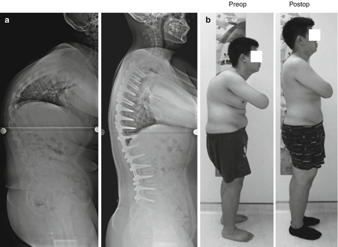

Fig. 7.7
(a) Lateral view of a 27-year-old male suffering ankylosing spondylitis related thoracic kyphosis. Postoperative lateral radiograph of spine showing correction of deformity with 2 SPOs at apex (T9–T12) and multiple POs above and below the apex. (b) Preoperative and postoperative clinical appearance
It is generally believed that an angular kyphosis, such as seen with posttraumatic kyphosis, is more amenable to a PSO or VCR. However, sometimes performing SPO and creating open wedges at and around the apex can obtained satisfactory correction with less neurological risk with the middle column functioning as the pivot point comparing with PSO or VCR (Fig. 7.8).
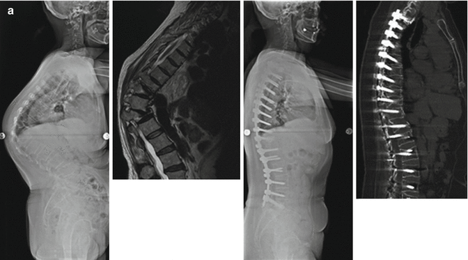
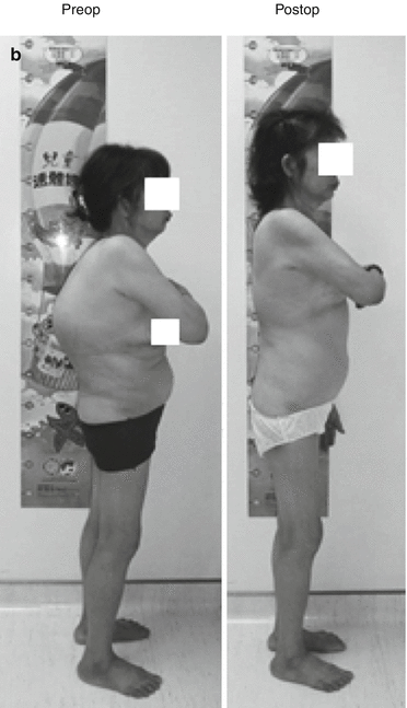


Fig. 7.8
It is generally believed that an angular kyphosis, such as seen with posttraumatic kyphosis is more amenable to a PSO or VCR. However, sometimes performing SPO and creating open wedges at and around the apex can obtained satisfactory correction with less neurological risk with the middle column functioning as the pivot point comparing with PSO or VCR. (a) Lateral view of a 50-year-old female suffering posttraumatic angular kyphosis. Postoperative lateral radiograph showing correction of the deformity with SPO at and around the apex (T10–T11 and T11–T12). (b) Preoperative and postoperative clinical appearance
Sagittal imbalance in a patient who has had a prior fusion to L4 with subsequent degeneration of L4–L5 and L5–S1 varies quite a bit from patient to patient. If osteotomy can be performed through the prior lumbar fusion segments, this is best treated with a PSO. On the other hand, if osteotomy cannot be performed through the prior lumbar fusion segments for any reason, then treat this by SPO and structural graft placement at L4–L5 and extension to the pelvis for structural support (Fig. 7.9).


Fig. 7.9


A 67-year-old female suffering postinstrumentation kyphoscoliosis. Osteotomy cannot be performed through the prior lumbar fusion segments. (a) Preoperative radiograph and CT showing T4–T12 kyphosis = 45°, T12–S1 lordosis = 9°, PI = 36°, PT = 40°, and SVA = 14 cm (b) Postoperative radiograph and CT showing correction of deformity with SPO at L4–L5 and POs at T9–T11, T4–T12 kyphosis = 13°, T12–S1 lordosis = 35°, PI = 36°, PT = 20°, and SVA = 1.7 cm. (c) Preoperative and postoperative clinical appearance
Stay updated, free articles. Join our Telegram channel

Full access? Get Clinical Tree


