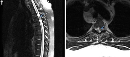
Figure 17.1
MRI scan showing intramedullary spinal cord tumor (blue arrows) on side view (sagittal, left image) and top view (axial, right image) of spine
Intradural Extramedullary Tumors
Tumors in this category arise from, or under, the outer layer of the spinal cord (dura), but external to the actual spinal cord (Fig. 17.2). Meningiomas and nerve sheath tumors (from the covering of the nerves) can develop in this compartment. Nerve sheath tumors include schwannomas and neurofibromas. Spinal meningiomas are most commonly in the thoracic spine, and grow very slow. Often times, they also have high calcium content [6]. Schwannomas and neurofibromas are benign nerve sheath tumors, but neurofibromas have the potential to become cancerous.


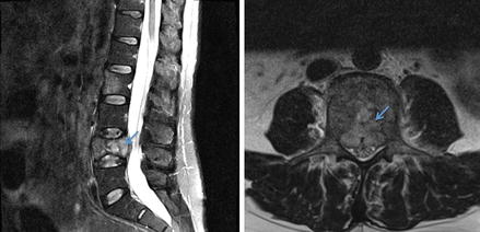
Figure 17.2
MRI scan showing intradural extramedullary spinal cord tumor (blue arrows) on side view (sagittal, left image) and top view (axial, right image) of spine
Extradural Tumors
Tumors in this category most often arise from the vertebral bodies (Fig. 17.3). As discussed, metastasis can occur anywhere in the spine, but will most often arise here, and are the most common extradural spine tumor. The most common metastasis in this area include prostate cancer, breast cancer, and lung cancer. This location is also home to several uncommon primary tumors. Tumors found here include: chordomas (rare bone tumors), sarcomas (usually in younger patients), lymphoma, plasmacytomas and multiple myeloma, and eosinophilic granuloma (Langerhans cell histiocytosis). Benign (nonmalignant) tumors in this category include: osteoid osteomas (found in long bones, <2 cm in size), osteoblastomas (>2 cm in size), osteochondromas (which has both bone and cartilage), chondroblastomas (arises from immature cartilage), giant-cell tumors, vertebral hemangiomas (composed of thing-walled blood vessels, and rarely cause symptoms), and aneurysmal bone cysts.


