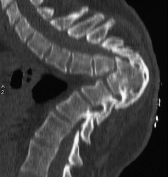
86 A 40-year-old woman with a history of back pain since adolescence complained of worsening pain but no neurologic deficit. Reconstructed computed tomography (CT) scan of the cervicothoracic spine is notable for extreme thoracic kyphosis and vertebral segmentation anomaly (Fig. 86-1). FIGURE 86-1 Dramatic thoracic kyphosis with segmentation anomaly. Congenital kyphotic deformity of the spine Surgical deformity correction Congenital kyphotic deformity of the spine encompasses a variety of spinal anomalies. Scoliosis with kyphosis or lordosis results from formation or segmentation failure. Failures of formation are hemivertebra and wedge vertebra, and segmentation failures include unilateral unsegmented bar and block vertebra. Over 50% of these patients have additional anomalies that can be related to the neuraxis (tethered cord, syringomyelia, or most commonly diastematomyelia), or related to other organs (musculoskeletal, genitourinary, or cardiovascular). Screening tests to look for associated abnormalities are strongly recommended and routine procedure prior to any surgical correction of these deformities. Congenital kyphotic deformities almost always involve the thoracic and thoracolumbar spine. Neurologic findings are more common if kyphosis has resulted from failure of formation. This produces a sharp angular gibbus compared with the smooth, round kyphosis seen in failure of segmentation. The presence of neurologic findings, curve progression, and patient age at time of presentation dictate treatment options. Children with kyphosis secondary to failure of formation are fused posteriorly in situ if there is less than 55 degrees of angulation. Additional anterior releases are necessary if the kyphosis is 55 degrees or greater.Attempting to correct a severe kyphosis carries a high risk of neurologic deficit. In the older patient with a milder deformity, particularly when related to segmentation failure, posterior fixation is adequate. The extracavitary approach can be used for patients with more dramatic corrective needs (e.g., decompression, vertebral osteotomy, and strut grafting), followed by posterior multilevel segmental fusion. Winter RB, Moe HH, Wang JF. Congenital kyphosis: its natural history and treatment as observed in a study of one hundred and thirty patients. J Bone Joint Surg Am 1973;55:223–256
Spinal Deformity
Presentation
Radiologic Findings
Diagnosis
Treatment
Discussion
SUGGESTED READING
< div class='tao-gold-member'>
Spinal Deformity
Only gold members can continue reading. Log In or Register to continue


Full access? Get Clinical Tree


