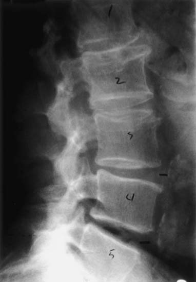
3 I. General considerations A. Spinal imaging modalities (Table 3–1) 1. Plain radiographs 2. Computed tomography 3. Magnetic resonance imaging (MRI) 4. Bone scintigraphy 5. Myelography 6. Angiography 7. Discography B. A thorough history and physical examination should lead to a preliminary clinical diagnosis that should predicate both the selection and timing of imaging tests. 1. Diagnostic tests should be used to confirm information ascertained during the history and physical examination. C. Selection of imaging tests should be based on the appreciation of the sensitivity, specificity, and accuracy of various imaging modalities in conjunction with different disease processes. 1. Acute neck or back pain and radiculopathy a. Natural history is that of improvement with conservative treatment b. Diagnostic imaging should be delayed until 4 to 6 weeks after the onset of symptoms. (1) Exceptions to earlier imaging evaluation include (a) Trauma (b) Progressive neurological deficit (c) If neoplasm or infection suspected 2. Imaging evaluation alone without clinical correlation is associated with an extremely high false-positive rate.
Spinal Imaging and Diagnostic Tests
♦ Imaging Modalities
| Imaging Modality | Indications/Advantages | Limitations |
| Plain radiographs |
|
|
| Computed tomography |
|
|
| Myelography and computed tomographic myelography |
|
|
| MRI |
|
|
| Bone scintigraphy (technetium99m, gallium-67 citrate, indium-11 White Blood Cell (WBC) scan) |
|
|
| Discography |
|
|


