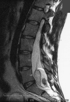
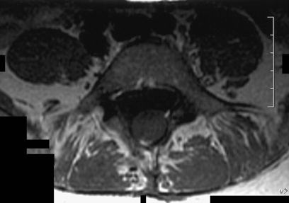
80 A 38-year-old woman had urinary retention. Workup found a large sacral teratoma, and she underwent a subtotal resection, with some clinical improvement. She had further medical treatment and pain management, but currently presents with worsening urinary symptoms and mild right lower extremity weakness. FIGURE 80-1 Sagittal view lumbosacral MRI shows a heterogenous intradural, tethering lesion. Recurrent teratoma is seen behind the L5-S1 segment on magnetic resonance imaging (MRI) (Figs. 80-1 and 80-2). A heterogeneous intradural lesion is observed tethering the spinal cord down to L5. Spina bifida is also noted. Recurrent teratoma. Specimen from the filum terminale is described as adipose tissue (Fig. 80-3). Specimen from the intradural mass is diagnosed as a mature teratoma with cuboidal and squamous elements and mature sebaceous sweat glands (Fig. 80-4). This patient had a redo lumbosacral laminectomy, adhesiolysis, and tethered cord release. This patient has a chronic cauda equina-type picture, with a recurrent, hard to treat lesion. Gross total surgical resection is recommended at the onset, because further surgical attempts carry a significant morbidity and are very challenging. In this case, her tethered cord must be released with lysis of adhesions, to alleviate her symptoms.
Spinal Teratoma
Presentation

Radiologic Findings
Pathology Findings and Diagnosis
Treatment
Discussion

Spinal Teratoma
Only gold members can continue reading. Log In or Register to continue

Full access? Get Clinical Tree


