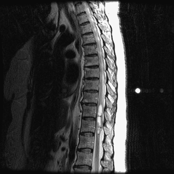
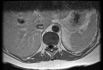
65 Asim Mahmood A 60-year-old woman had gait difficulty and back pain. On examination, her reflexes were brisk and she had lost proprioception. Her symptoms were worse at night. An intramedullary lesion with an associated syrinx is noted on thoracic magnetic resonance imaging (MRI) (Figs. 65-1 and 65-2). FIGURE 65-1 Sagittal MRI is notable for a thoracic intramedullary lesion with an associated syrinx. Pathology was diagnostic for a thoracic ependymoma A thoracic laminectomy for lesion excision with somatosensory evoked potential (SSEP) monitoring was done. Intraoperative photos display the tumor and the resection bed (Figs. 65-3 and 65-4). Ependymomas are glial tumors that arise from ependymal cells. In the spine, they usually affect adults. The most common spinal variant is the myxopapillary ependymoma of the conus and cauda equina. An important intraoperative difference between the other more commonly seen intramedullary spinal tumor—the astrocytoma—is that the plane between the ependymoma and normal cord is well developed. Recurrence in the spine after a gross total resection is rare. The need for adjuvant treatment depends on tumor type and the totality of resection.
Spinal Tumor
Presentation
Radiologic Findings

Diagnosis
Treatment
Discussion

Spinal Tumor
Only gold members can continue reading. Log In or Register to continue

Full access? Get Clinical Tree


