♦ Preoperative
Operative Planning
- Make sure epileptologist and EEG technologist are present to interpret electrocorticography (ECoG)
- Determine if additional subdural strip and/or grid electrodes are necessary
- Order necessary electrodes prior to surgery
- Platinum electrodes are more compatible with MRI
- Stainless steel electrodes are less expensive
- Platinum electrodes are more compatible with MRI
- Ensure that appropriate connecting cables and blocks are available
- Frameless or frame-based stereotaxy is required
- Ensure that equipment is compatible with diameter of electrodes
- May require a special cannula and/or reducing sleeve
- Ensure that equipment is compatible with diameter of electrodes
- Obtain stereotactic planning gadolinium-enhanced MRI prior to surgery
- Contrast is administered for visualization of the vasculature
Equipment
- Electrodes
- Neuronavigation system
- EEG recording device/computer
- Slotted cannula for electrode placement
- Stereotactic biopsy tray; lockable arm to hold the cannula in place
- Reducing sleeve if necessary
Anesthesia Issues
- Intravenous cefazolin (1 g) is given prior to the procedure
- Foley catheter
- Arterial line
- Minimize anesthesia during the ECoG
- Avoid high concentrations of halogenated inhalational anesthetics
- Avoid barbiturates, benzodiazepines, nitrous oxide, or propofol
- We use a combination of fentanyl and isoflurane < 0.2%.
- Mannitol is avoided to reduce brain shift
- Minimize anesthesia during the ECoG
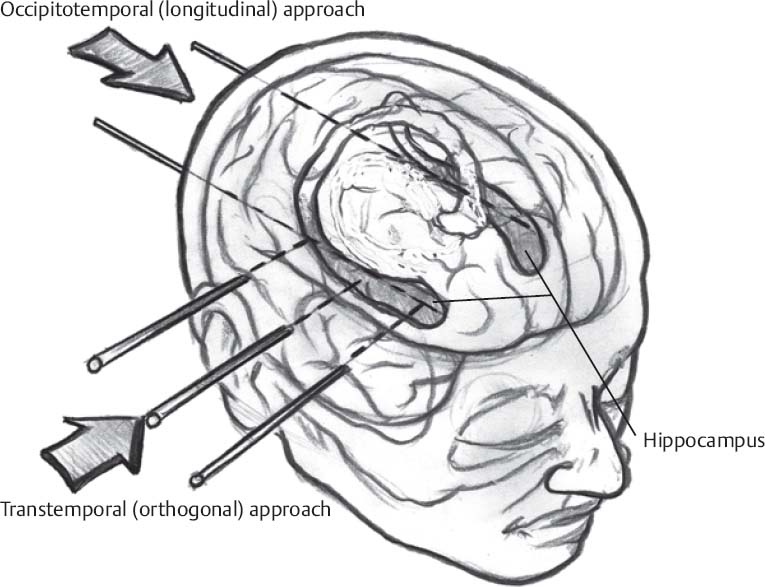
♦ Intraoperative (Fig. 59.1)
Positioning
- For unilateral temporal depth electrode placement in combination with subdural grids, a lateral temporal route is used.
- The patient is in the supine position with the head turned away from the side of interest.
- A shoulder roll is usually necessary.
- The patient is in the supine position with the head turned away from the side of interest.
- For bilateral temporal depth electrode implantation, the occipitotemporal approach is employed.
- The patient is in a semisitting position with the neck flexed.
- For both positions, the head is placed in a three-pin Mayfield frame, which is attached to the operating table.

Only gold members can continue reading. Log In or Register to continue


