Clinical Syndromes
History/Examination
Because of the anatomy of the cerebral vasculature, thromboembolic stroke tends to result in particular sets of symptoms and signs or ‘stroke syndromes’. Many of these are uncommon or rare (examples can be reviewed at the Internet Stroke Center www.strokecenter.org/prof/syndromes/). For more common stroke types, the OCSP classified strokes into broad syndromes to aid prediction of prognosis. Though more than 20 years old, these syndromes are still useful; they are described below. Anterior circulation refers to the territory of an internal carotid artery—middle cerebral artery and/or anterior cerebral artery. Posterior circulation refers to the territory of the vertebral and basilar arteries.
- Total anterior circulation stroke (TACS): Results from occlusion of the internal carotid artery or proximal middle cerebral artery:
 Contralateral hemiparesis with or without sensory deficit involving two or more out of three body areas (face/upper limb/lower limb) opposite the lesion.
 Homonymous visual field defect opposite the lesion.
 Higher cortical dysfunction (dysphasia, neglect, visuospatial problems, depending on the hemisphere affected and cerebral dominance).
- Partial anterior circulation stroke (PACS): Results from occlusion of a branch of the middle cerebral artery. The PACI syndrome reflects less extensive neuronal loss than the TACI syndrome, comprising two of the three deficits listed above or as follows:
 Isolated higher cortical dysfunction (i.e. isolated dysphasia or predominantly proprioceptive deficit in one limb), or
 Motor/sensory deficits restricted to one part of the body (face/arm/leg contralateral to the lesion).
- Posterior circulation stroke (POCS):
 Isolated (or occasionally bilateral) homonymous haemianopia due to occipital cortex damage.
 Brainstem ischaemia with cranial nerve involvement ipsilateral to the lesion (diplopia, vertigo, dysphonia and dysarthria) and/or various patterns of ophthalmoplegia, as well as ipsilateral long-tract motor signs and contralateral sensory signs (e.g. the lateral medullary syndrome or Wallenberg syndrome).
 Cerebellar ischaemia with ipsilateral limb (cerebellar hemispheres) and trunk (midline cerebellum) ataxia, vomiting, vertigo with horizontal nystagmus, and usually mild ipsilateral limb weakness.
 Midbrain ischaemia with a variety of possible signs of both anterior and posterior circulation syndromes, with or without somnolence or behavioural disturbance.
- Lacunar stroke (LACS): Results from in situ thrombosis of small end arteries to basal ganglia and internal capsule or of small arteries in the ‘watershed zones’ between vascular territories. Hypertensive, diabetic patients are particularly at risk.
 Pure motor: A unilateral motor deficit (internal capsule or pons).
 Pure sensory: Similar to motor in distribution, proprioception is spared (thalamus).
 Sensorimotor: Combined deficit (thalamus and internal capsule).
Many others exist, for example ataxic hemiparesis, dysarthria clumsy hand syndrome.
Investigations
See Chapter 13.
Management
Treatment follows the following principles:
- Restoration of blood flow: Usually thrombolysis, possible only in some carefully selected patients.
- Prevention and treatment of complications: For example aspiration pneumonia, thromboembolism, spasticity, depression and seizures.
- Rehabilitation: Physical, occupational, vocational, speech therapy and neuropsychological rehabilitation.
- Secondary prevention: For example antiplatelet and anticoagulant therapy, treatment of hypercholesterolaemia and treatment of hypertension.
- Long-term support: (beyond the scope of this chapter).
Stroke Units
Stroke units should be the place of care for stroke patients. Staffed with multidisciplinary stroke specialists, they reduce the relative risk of patient death by about 15% and institutional care by about 20%. A stroke unit is more effective than either a stroke bay on a medical ward or a mobile stroke team.
Thrombolysis
The clot dissolving drug recombinant tissue plasminogen activator (rt-PA), and possibly others (though not streptokinase), has been shown to improve outcome in ischaemic stroke in carefully selected patients. Unfortunately, the majority of patients are not eligible for treatment, either because they attend hospital too long after the onset of symptoms or because they have another contraindication to treatment.
The following are the important facts related to recombinant tissue plasminogen activator given intravenously within 3–4½ h:
- Is more effective the earlier it is given.
- Reduces frequency of death or dependency at around 6 months by about one-third (relative risk) on average.
- Increases early death due to intracranial haemorrhage by approximately three–four times.
- Is more likely to cause haemorrhage the later it is given.
Alternatives to intravenous thrombolysis include the following:
- Intra-arterial thrombolysis: This appears to be effective up to 6 h after the onset of symptoms.
- Intra-arterial clot extraction: This appears to be the most effective method for restoring blood flow, but its effectiveness has not been proven in terms of clinical outcomes. Unlike intravenous thrombolysis, it can be attempted in patients taking oral anticoagulation.
- Combinations of intravenous thrombolysis and the above treatments.
Aspirin
A dose of 300 mg of aspirin daily given as soon as possible after stroke for 14 days improves outcomes at 6 months and reduces recurrent ischaemic strokes within 14 days (absolute risk reduction 1.1% and relative risk reduction 28% in the International Stroke Trial).
Medical Management: Secondary Prevention
Standard medical management for secondary prevention of thromboembolic stroke in patients without atrial fibrillation is as follows:
- Rule out intracerebral haemorrhage, for example with brain CT.
- Aspirin 300 mg od immediately (clopidogrel 75 mg od if aspirin intolerant).
- Change to combination of aspirin 75 mg od + dipyridamole MR 200 mg bd after perhaps 2 weeks (clopidogrel 75 mg od alone if either drug not tolerated).
For all patients with thromboembolic stroke:
- If cholesterol is ≥3.6, start simvastatin 40 mg nocte (MRC Heart Protection Study). Other agents, for example fibrates, may be used in patients unable to tolerate statins. Because of the potential risk of haemorrhage associated with statin use, NICE does not recommend immediate statin use (e.g. wait 72 h from stroke before starting statins).
- If systolic BP is ≥110 mmHg, start antihypertensives, aiming for treatment with an ACE inhibitor and a thiazide diuretic (best evidence is the PROGRESS trial, in which perindopril 4 mg daily plus indapamide 2.5 mg daily reduced relative stroke risk by 43% compared to placebo). The role of acute blood pressure lowering after stroke remains unclear.
Further antiplatelet options:
- As suggested by the EXPRESS trial, in patients with minor stroke without atrial fibrillation in whom treatment can be started within 48 h (i.e. within the highest risk period), it may be reasonable to give aspirin 75 mg plus clopidogrel 75 mg (after a 300 mg loading dose of aspirin) for a month, followed by aspirin 75 mg plus dipyridamole MR 200 mg bd.
Further cholesterol-lowering treatment:
- Treatment should be increased for patients who fail to reach a target of 4 mmol/L total cholesterol or 2 mmol/L LDL cholesterol (NICE guideline). High doses of statins (atorvastatin 80 mg) may increase the rate of intracerebral haemorrhage following stroke (SPARCL study).
- Further antihypertensives.
- Treatment should be further increased if a target blood pressure of 140 mmHg systolic and 90 mmHg diastolic is not met (NICE guideline).
Atrial fibrillation:
- Patients with atrial fibrillation should be considered for anticoagulation. In patients with stroke, there is an increased risk of intra-cerebral haemorrhage with all anticoagulants, therefore a period (usually 2 weeks) should be left between the stroke and starting anticoagulant medication. For further principles of anticoagulant management in atrial fibrillation, see Chapter 2.
Nutrition and feeding Dysphagia with aspiration is common in stroke, though dysphagia usually resolves in surviving patients. The FOOD series of clinical trials found the following:
- Early nasogastric feeding appears to improve survival, but this may be at the expense of increased dependent survival.
- Percutaneous endoscopic gastrostomy (PEG) should be reserved for patients in whom nasogastric feeding cannot be used or who require prolonged feeding.
- Routine nutritional supplementation does not improve outcomes.
Thromboprophylaxis Deep venous thrombosis with or without pulmonary embolism is common following stroke, particularly in hemiplegic and non-ambulant patients. All anticoagulants increase the risk of haemorrhage following stroke; in the acute phase, this generally exceeds any benefit. However, thromboprophylaxis with low-molecular weight heparin may be beneficial after the acute period, in patients with low risk of bleeding (e.g. no haemorrhagic transformation of the cerebral infarct) and high risk of thromboembolism. (e.g. severe immobility/hemiplegia, dehydration, previous venous thromboembolism and comorbidity such as cancer).
Carotid endarterectomy and stenting Carotid endarterectomy may be indicated for patients following a stroke if they have shown a reasonable recovery (reflecting a reasonable capacity to benefit from the procedure). For further details, see Chapter 13.
Complications
Complications after stroke include the following:
- Aspiration pneumonia: Dysphagia after stroke puts patients at risk of aspiration and therefore pneumonia.
- Venous thromboembolism: Deep venous thrombosis (DVT) is common after stroke, and pulmonary embolism (PE) is not infrequent. Severe weakness and immobility put patients at high risk. The CLOTS trial showed no benefit of thigh-length compression stockings in stroke patients. Anticoagulants used to prevent and treat DVT and PE can cause bleeding into damaged brain, therefore their use often involves a difficult risk/benefit assessment (see above).
- Depression: Depression is common after stroke. Antidepressants can be beneficial. Psychological therapies may also have benefit.
- Spasticity: Spasticity, or increased muscle tone, after stroke can be painful and interfere with function. Physiotherapy, antispasmodic medication, such as baclofen, and botulinum toxin are the main treatments. Intrathecal baclofen and surgery for contractures are less commonly used.
- Seizures: Seizures may occur early (within 2 weeks, about half within 24 h) or late (after 2 weeks) after stroke. Late seizures have a higher rate of recurrence; about one-third of patients with early seizures and one-half with late seizures go on to epilepsy.
Note on Rehabilitation
Rehabilitation should begin soon after stroke, though it is not clear exactly how early this should happen. Physiotherapy improves gait and aims to restore other aspects of physical function, while reducing risk of falls. Occupational therapy aims to improve function by, for example, simplifying and practising tasks. Speech and language therapy aims to improve speech production, intelligibility and comprehension and swallowing ability. Speech and language therapists regularly assess aspiration risk to guide decisions about route of feeding. Rehabilitation goals should be set and their attainment regularly reviewed. More detailed discussion of stroke rehabilitation is beyond the scope of this chapter.
Prognosis
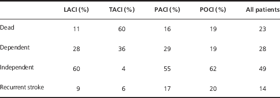
The natural history of the major subtypes of thromboembolic stroke was investigated on a population scale in the Oxford Community Stroke Project (1981–1984). Table 14.2 shows prognosis at 1 year in terms of outcome and recurrent stroke.
Table 14.2 Natural history of stroke at 1 year (data from the Oxford Community Stroke Project)
Differential Diagnoses (Also See Table 13.2)
The differential diagnosis of thromboembolic stroke is similar to that of TIA, though there are differences (Table 14.3).
Table 14.3 Differential diagnoses for thromboembolic stroke with points to note.
| Clinical conditions | Points to note |
| Subdural haematoma (Chapter 33) | This is not conventionally classed as a stroke. Brain scans should be examined closely as small subdural haematomas are easily missed |
| Subarachnoid haemorrhage (Chapter 16) | Subarachnoid haemorrhage is sometimes classified as a type of stroke. Along with the typical sudden onset of severe headache, it can cause focal neurological deficits |
| Migraine (Chapter 12) | Migraine aura results from temporary cerebral dysfunction. It generally does not last more than 24 h but sometimes can. Migraine with aura may itself occasionally cause stroke |
| Previous stroke with intercurrent illness | Patients with cerebral damage from previous stroke frequently have recurrence of their stroke symptoms when they are systemically unwell, for example due to infection |
| Epileptic seizure (Chapter 17) | Though Todd’s paresis (weakness persisting after a seizure) does not generally last 24 h, in patients with ongoing recurrent focal seizures or in patients with severe generalised seizures and previous focal brain damage, seizures may mimic stroke |
| Functional symptoms | Symptoms, especially hemiparesis, in the absence of organic illness are surprisingly common. The duration of symptoms is variable |
| Vestibular disorders (Chapter 8) | Vestibular disease can cause vertigo of abrupt onset and mimic vertebrobasilar stroke |
| Pressure palsies, for example radial nerve palsy | These can usually be differentiated in the history but can cause difficulty in patients with multiple vascular risk factors, particularly as acute stroke often presents with flaccid tone |
| Hypoglycaemia | Hypoglycaemia can precipitate stroke but will not usually cause symptoms for more than 24 h duration |
< div class='tao-gold-member'>


