Remember: CT head is the initial diagnostic investigation of choice in ICH.
- Routine blood tests FBC (including platelet count), U & E (check for evidence of end organ damage related to hypertension and hyponatraemia related to SIADH), LFT, G & S, clotting profile (PT, APTT, and INR).
- ECG Look for evidence of left ventricular hypertrophy.
- Imaging
 CT: Initial diagnostic investigation of choice, can be done rapidly and shows blood as high density (brighter) compared to brain parenchyma immediately after the event, shows degree of midline shift and mass effect, presence of associated intraventricular extension with or without hydrocephalus (Figure 15.1).
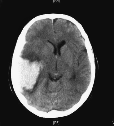
 MRI: Useful at a delayed interval for demonstrating underlying structural abnormalities, for example a tumour.
 Cerebral angiography: Generally recommended unless patients are above 45 years of age with known hypertension and have haemorrhage in putamen or thalamus. An angiogram should not delay the initial emergency treatment if required and can be performed at a delayed interval to increase the diagnostic yield.
Figure 15.1 CT demonstrates ICH in right temporal lobe with mass effect and effacement of ipsilateral frontal horn of right lateral ventricle.
Only gold members can continue reading. Log In or Register to continue


