Remember: SAH should be strongly suspected in any patient describing a severe headache of instantaneous onset even in the absence of other symptoms or signs of meningism.
Other symptoms:
- Irritation of lumbar nerve roots by SAH may cause lumbar back pain.
- Compression of third nerve by an enlarging aneurysm may cause diplopia and a ptosis.
Examination
Depressed level of consciousness/coma:
- Patients may be comatose from the outset or have a depressed level of consciousness.
- Document the Glasgow Coma Score (GCS).
- Grading by WFNS grade (Table 16.1).
Table 16.1 WFNS grading of subarachnoid haemorrhage.
| Grade | GCS | Focal neurological deficit |
| I | 15 | Absent |
| II | 13–14 | Absent |
| III | 13–14 | Present |
| IV | 7–12 | Present or absent |
| V | <7 | Present or absent |
Focal neurological deficits:
- Due to compression of adjacent neural structures by either the expanding aneurysm or an associated intracerebral haematoma.
- Basilar or posterior communicating artery aneurysm expansion may cause a IIIrd nerve palsy.
- Intracavernous sinus carotid aneurysms may present with opthalmoplegia (III, IV and VI nerves) and facial pain (V nerve, ophthalmic division).
- Internal carotid or anterior communicating artery aneurysms may compress the chiasm or optic nerves causing visual field deficits and the pituitary stalk causing hypopituitarism.
- Middle cerebral artery aneurysm may rupture into the parenchyma causing an intracerebral haematoma that compresses or damages the motor cortex or internal capsule causing a hemiparesis.
Meningism:
- Neck stiffness, ‘nuchal rigidity’ present on passive flexion of neck.
- Positive Kernig’s sign (flex thigh with patient supine, then extend the knee, positive if causes pain in hamstrings preventing full extension). Usually seen >6 h following bleed.
Ocular haemorrhage:
- Seen on fundoscopy, often bilaterally.
- Preretinal (subhyaloid) haemorrhage, seen as blood obscuring retinal vessels in the region of optic disc, and/or vitreous haemorrhage (Terson’s syndrome), seen as vitreous opacity.
- Occurs in approximately 10–20% of bleeds.
- In most cases, bleeding clears spontaneously but can lead to permanent visual loss.
Fever:
- Common.
- May be due to impairment of heat regulation due to hypothalamic damage or secondary infection.
Cardiovascular signs:
- Hypertension may be present.
- Cardiac arrhythmias may occur.
- In obtunded patients with raised ICP, Cushing’s response (hypertension and bradycardia) may be seen.
- If a cardiac murmur is present on auscultation, consider infective endocarditis as a possible cause of SAH (mycotic aneurysm).
Investigations
Investigations are performed firstly to establish the diagnosis of SAH and then to identify the cause of the SAH.
To establish diagnosis:
CT head:
Remember: CT head is the initial investigation of choice in patients with suspected SAH.
- Initial investigation if SAH is suspected following clinical evaluation.
- Will detect 95% of cases if done within 48 h of onset of symptoms.
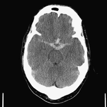
- Location of the blood can localise the likely location of the aneurysm (Figure 16.1).
- Look also for complications of SAH, including intracerebral haematoma, intraventricular haemorrhage, cerebral infarction and hydrocephalus.
- Rarer causes of SAH such as tumours or AVMs may be seen.
Figure 16.1 Axial CT Head showing acute subarachnoid blood (white) in right sylvian fissure, interhemispheric fissure and basal cisterns following rupture of an anterior communicating artery aneurysm.
Only gold members can continue reading. Log In or Register to continue


