Fig. 3.1
Lateral extensions of the dura, myelography with coronal and axial reformats (a, b) and axial T2w (c). The lateral extensions of the dura are the dural sleeves containing the nerve roots as well as CSF thus allowing assessment of the root sleeves on MRI (c) and myelography (a, b)
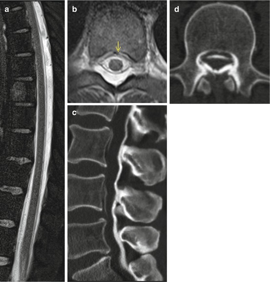
Fig. 3.2
Epidural space. The epidural space normally shows a fat-isointense signal on MRI imaging. In this case, the epidural space shows a water-isointense signal on fat-saturated T2w due to CSF leakage which caused intracranial hypotension (arrow) (a, b). An epidural blood patch was performed to seal the leakage (c, d). CT shows the epidural distribution of the applied hyperdense mixture of blood and contrast media. The undulating appearance of the epidural space on the sagittal reformations (d) is due to respiratory motion artifacts. Axial CT (c) and sagittal reformations (d)
Imaging: The dura can be detected on MRI as a thin black line on T2w. After the application of contrast agent, an enhancement can be seen due to a lack of endothelial tight junctions of the vessels.
The arachnoid is attached to the dura, follows its contours and also extends within the dural sleeves. It forms a fine network of connective tissue fibres that extend to the innermost layer of the meninges. An incomplete longitudinal midline membrane, which connects the pia and the cord dorsally to the dura, is called septum posticum and can be found in the cervical and the upper thoracic spine. It partially divides the subarachnoid space.
The pia is the innermost and thinnest layer of the meninges and is adherent to the surface of the cord and spinal nerve roots. It also enters the dural sleeves where it fuses with the arachnoid. Caudally it covers the filum terminale. Pia and arachnoid are also called leptomeninges.
The denticulate ligament exists between level C1 and L1 and consists of flat fibrous perforated sheets that support the spinal cord. It extends laterally from the pia between ventral and dorsal roots, inserts into the dura mater and therefore partially separates the subarachnoid space in an anterior and a posterior part. There are 20–22 pairs of denticulate ligaments.
3.2 Spaces
The real and potential spaces of the vertebral canal are located between the meningeal layers or adjacent structures. A potential space is only present postmortem or due to pathologic conditions, such as haemorrhage.
Related posts:


Stay updated, free articles. Join our Telegram channel

Full access? Get Clinical Tree


