(1)
Marina Spine Center, Marina del Rey, CA, USA
1.
Position the patient supine. Slightly hyperextend the neck, rotated away from the side of the approach. Use the inflatable cervical pillow for support; a small roll under the shoulder often helps to extend the neck. Location of the thoracic duct and recurrent laryngeal nerve becomes even more important at this level. Approaches from the left for C6 through T2 are directly in the vicinity of the thoracic duct. Identify the thoracic duct when possible and protect it. A large fatty meal the day before surgery will help. If the thoracic duct is inadvertently divided, double ligate both ends well. The approach to the right should definitely identify the recurrent laryngeal nerve and protect it. We recommend the right supraclavicular approach with identification of the recurrent laryngeal nerve.
2.
Make a transverse incision approximately one fingerbreadth above the clavicle from the midline to the posterior border of the sternocleidomastoid muscle (see Fig. 9.1). After division of the skin and subcutaneous tissue and the placement of small skin self-retaining retractors, incise the platysma muscle in the line of the incision. As with the higher approaches, identification of the medial border of the sternocleidomastoid is imperative.
3.
In addition, identify and define the anterior and posterior borders of the sternocleidomastoid muscle. The external jugular vein, although somewhat variable, is usually directly in the operative field and the anterior jugular vein is positioned more medially. Divide if necessary.
4.
Incise the external investing fascia. Pass a probe or finger laterally from the medial border of the sternocleidomastoid to clear off the venous structures underneath the clavicular head of the sternocleidomastoid.
5.
Divide the sternocleidomastoid laterally to medially, watching for the internal jugular vein [1]. If required for visualization, remove the sternal head of the sternocleidomastoid muscle in the same fashion. Eventual reattachment depends on suturing the fascial coverings of the muscle.
6.
Retract the divided sternocleidomastoid in a cephalad-caudad direction with self-retaining blunt retractors. The floor of the incision, at this point, consists of the middle cervical fascia, which contains the omohyoid and the sternohyoid muscles (see Fig. 7.3).
7.
Get Clinical Tree app for offline access

Enter the middle cervical fascia lateral to the carotid sheath. Bluntly dissect to the surface of the anterior scalene muscle (Fig. 10.1). The superficial surfaces of the anterior scalene are composed of the outer layer of the prevertebral fascia, the third and deepest of the fascial layers addressed in this approach. Lying on the surface of the anterior scalene is the phrenic nerve. The phrenic nerve crosses lateral to medial, cephalad to caudal (Figs. 10.2, 10.3). Retract the phrenic nerve medially after freeing it from the surface of the anterior scalene muscle. Identify the large internal jugular vein medially and feel the carotid pulse (Fig. 10.4). Although there are standard approaches to retract the carotid sheath laterally [2] (Fig. 10.5), attempt to retract the internal jugular vein and carotid sheaths medially (Figs. 10.1, 10.6). Retract the phrenic nerve to obtain good visualization of the anterior scalene muscle. The brachial plexus courses under the anterior scalene, between it and the middle scalene. The brachial plexus and suprascapular nerves are more superficial at the lateral border of the anterior scalene (Fig. 10.3).
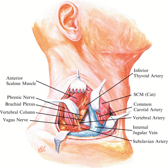
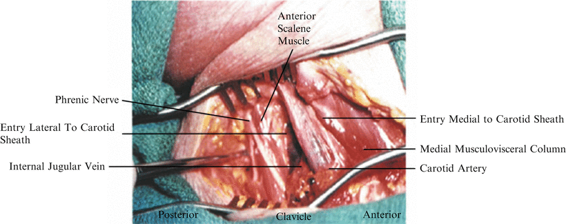
Fig. 10.1
After a transverse incision approximately one fingerbreadth above the clavicle from the midline to the posterior border of the sternocleidomastoid muscle, as described in Staicnc Muscle, as described in Chapter 7, delineate the medial and lateral borders of the sternocleidomastoid muscle (SCM). Divide the sternocleidomastoid muscle, avoiding damage to the internal jugular vein and muscular venous branches under the sternocleidomastoid, by passing a peon or angled retractor medial to lateral under the muscle before cutting with electrocautery. This composite picture shows the cut and retracted sternocleidomastoid muscle, the laterally retracted anterior scalene muscle and phrenic nerve, and a medially retracted internal jugular vein
Fig. 10.2


After division and retraction of the sternocleidomastoid muscle, three basic areas of approach are seen. (1) By retracting the phrenic nerve and resecting the anterior scalene muscle approach from the most lateral direction medially, an approach standardly used for a sympathectomy. (2) Lateral to the carotid sheath and medial to the anterior scalene muscle is an approach that allows medial retraction of the internal jugular vein and carotid sheath. (3) A more conventional approach medial to the carotid sheath and internal jugular vein may require division and ligation of the inferior thyroid artery as described in Chapters 6 and 10
Related posts:

Stay updated, free articles. Join our Telegram channel
Full access? Get Clinical Tree