Chapter 19 Sural Nerve
Anatomy
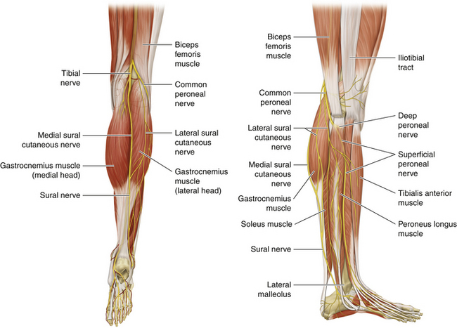
• The sural nerve is formed from two components derived from the tibial and peroneal nerves (Figure 19-1). The branching of the tibial and peroneal components and the main nerve is variable.
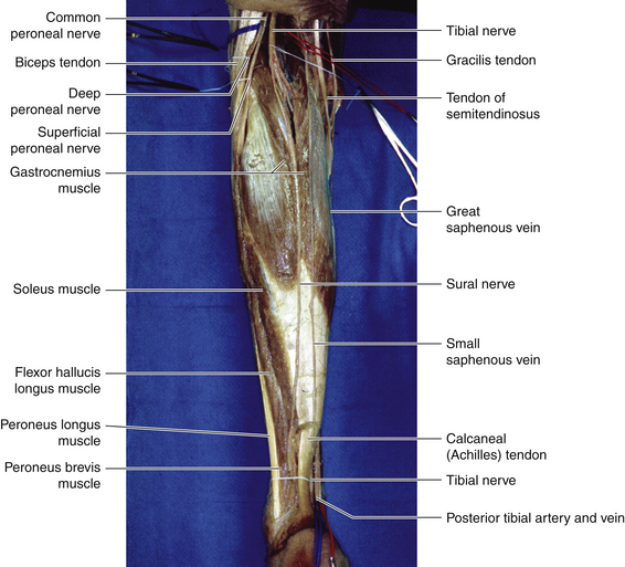
• The tibial nerve gives a single cutaneous branch—the sural nerve—at about the midline of the popliteal fossa. The sural nerve descends between the two heads of the gastrocnemius, deep to the investing fascia (Figure 19-2).
• Leaving the popliteal fossa at the inferior angle, the sural nerve descends in the groove between the two heads of the gastrocnemius and pierces the deep fascia proximally in the leg to join the lateral sural nerve.
• The level at which the various elements of the sural nerve pierce the investing fascia of the leg to run in the subcutaneous fat is variable.
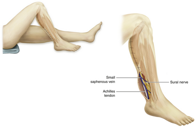
• Accompanied by the small saphenous vein, the sural nerve descends lateral to the Achilles tendon to the region located between the lateral malleolus and the area in front of the Achilles tendon (Figure 19-3). The point of junction of the two component nerves varies from immediately proximal to the lateral malleolus to the popliteal fossa. The specific pattern of a patient’s sural nerve cannot be forecast preoperatively.
• Distal to the lateral malleolus, the sural nerve runs along the lateral border of the foot and ends at the lateral side of the little toe. It supplies the posterior and lateral skin of the distal third of the leg and over the lateral border of the foot up to the tip of the little toe.

Figure 19-1 Posterior and lateral views of the lower extremity depicting the nerves and musculature.
Surgery
Patient Positioning
• When the patient is prone, as for an approach to the sciatic nerve or a posterior approach to the radial or axillary nerves, sural nerve exposure is easily accomplished, because the calves are uppermost.
• With the patient supine, as is the case for many nerve dissections, sural exposure is more difficult.
• A folded towel is clipped to the surface of the operating table so that the knee can later be flexed and the hip internally rotated, with the foot braced against the folded towel.
• A folded sheet under the buttock helps the surgeon internally rotate the leg a bit to expose the calf.
• The plan to harvest sural grafts should be rehearsed before the main operative site and sural donor sites are prepared and draped. This is particularly important if an over-the-patient instrument table is used, or if the operating room nurse changes the site of command halfway through the procedure.
Stay updated, free articles. Join our Telegram channel

Full access? Get Clinical Tree


