The treatment of patients with spinal disease has traditionally represented a large portion of general neurosurgical practice. Despite the fact that spinal operative procedures comprise the majority of the neurosurgical procedures performed in this country, the scientific foundation in support of their application and utility is relatively diminutive. Recently, a concerted effort has been made to scientifically analyze and evaluate neurosurgical practices in the assessment, diagnosis, prognosis, and treatment of patients with spinal disease. In this chapter, we provide examples of the application of critical evidence-based medicine methods to these aspects of the neurosurgical care of patients with spinal disease. The importance of this type of work, this method of critical thinking, the proper design of clinical studies to provide robust, meaningful medical evidence is paramount to our discipline. Neurosurgeons must critically evaluate and substantiate our practices and procedures for the benefit of our patients, our heralded profession, and ourselves. The assessment of patients with spinal disorders involves a clinical evaluation, including a neurological examination. The best evidence in the medical literature on the assessment of patients with spinal disease is found within the acute spinal and spinal cord injury literature. The assessment of patients who have sustained an acute spinal cord injury involves a neurological examination to document existing neurological function, and a functional outcome assessment to provide prognostic information about future functional skills and disabilities. A variety of scales and scoring systems has been proposed for each of these components of the neurological assessment, each with variable reliability and reproducibility. In 2002, a multitopic, 22-chapter medical evidence–based review on issues related to acute spinal trauma was completed, critically reviewed, and published.1 The Guidelines on the Management of Acute Cervical Spine and Spinal Cord Injuries includes a comprehensive chapter on neurological assessment. The Guidelines’ author group employed standard, stringent clinical epidemiology criteria in their review of the English-language literature on this topic. In addition, Hadley, Walters, et al1 developed benchmarks for the strength of the medical evidence on this subject.  7
7 
Surgery for Cervical Spine Trauma
David A. Vincent, Paul C. McCormick, Mark N. Hadley
Patient Assessment
Neurological Function Assessment
| Scale/Year | Description | Type | Notes |
|---|---|---|---|
| Frankel Scale91 (1969) | First stratified neurological scale for SCI patients. | Five-grade scale, A–E | Easy to use. Broad categorizations. Imprecise. Poor sensitivity to change, especially in groups C and D. No kappa values reported. |
| Yale Scale88 (1981) | Adaptation of British Medical Research Council’s. gradation | 10 selected muscle groups, graded 0–5. Two-point sensory scale for superficial pain, position sense, and deep pain. | Difficult to use at bedside. Not specific Bladder/bowel function not. considered. No kappa values reported. |
| ASIA Scale84 (1984) | American Spinal Injury Association created standards for the neurological classification of spinal injury patients | 10 muscle group scoring, 0–5 grades. Used Frankel scale as functional tool. | No scoring for sensory exam. Insensitive. No kappa values reported. |
| NASCIS Scale87 (1985) | Motor/sensory assessment employed in NASCIS trials I and II | 14 muscle groups, graded 0–5. Sensory (light touch and pin prick) scored for C2-S5 dermatomes, 0–3 points. | No functional abilities assessment. Separate scores for right and left side of body. |
| ASIA Scale89 (1989) | ASIA 1984 with refinements, addition of Sensory scoring (0–2 points) | More precise. kappa values reported: 0.50 to 0.67. | |
| ASIA ISCSCI92 (1992) | Revision of ASIA 1989 scale by American Spinal Injury Association in conjunction with International Medical Society of Paraplegia | ASIA 1989 with addition of motor index scores and the ASIA impairment scale. Incorporated FIM as functional assessment tool. | Improved sensitivity, especially with respect to functional assessment. Kappa values from 0.0 to 0.89 in several studies. Greatest variability in patients with incomplete injuries. |
| ASIA ISCSCI96 (1996) | Revision of ASIA IMSOP 1992 scale | No reports of intra- or interoperative server reliability yet published. |
Abbreviations: ASIA, American Spinal Injury Association; FIM, function independence measure; ISCSCI, International Standards for Neurological and Functional Classification of Spinal Cord Injury; NASCIS, National Acute Spinal Cord Injury Studies; SCI, spinal cord injury
Neurological examination and functional outcome assessment scales must be easy to use and apply, must appropriately characterize the patient’s neurological circumstances, and must be reliable from observer to observer and by the same observer on different occasions. Inter- and intraobserver reproducibility and accuracy must be high. They are best evaluated using a statistic called kappa (K) value and employing a Bayesian comparison. The authors determined that an assessment scale with an interobserver reliability kappa value of 0.81 or greater represented “near perfect” agreement between observers.2,3 A kappa value of 0.61 to 0.80 represented “substantial” agreement and a kappa value 0.41 to 0.60 represented “moderate” agreement. This ranking of reliability performance was translated into strength of recommendations for clinical application.
Employing these criteria, the Guidelines’ authors could not identify a single neurological examination scale with a kappa value of 0.61 or higher (Table 7–1). The American Spinal Injury Association/International Medical Society of Paraplegia (ASIA/IMSOP) Standards for Neurological and Functional Classification of Spinal Cord Injury published in 19964 appears to be the most accurate and reproducible neurological examination scale, but studies comparing interobserver reliability had not been published at the time of the Guidelines’ completion. At present, class III medical-evidence supports the use of the ASIA/IMSOP standard scale as an Option-level recommendation (Table 7–2).
The functional impairment measure (FIM) is the most reliable functional outcome assessment scale reported in the medical literature.4–15 According to the same rigidly applied assessment criteria, interobserver reliability for FIM has a documented kappa value of 0.76.9,13 It is considered to have class II medical evidence in support of a Guideline level recommendation for its use.
| Reference | Description | Evidence Class |
|---|---|---|
| Jonsson et al, 200078 | A study of the interrater reliability of the ASIA ISCSCI-92. Physicians and physiotherapists classified 23 patients according to the ISCSCI-92 and calculated kappa values. | III |
| Cohen et al 1996104 (S28–18) | A test of the ASIA ISCSCI-92. Participants completed a pretest and posttest in which they classified two patients who had an ASCI. | III |
| El Masry et al, 199679 | A study to assess the reliability of the ASIA and NASCIS motor scores. The motor scores of 62 consecutive ASCI patients were retrospectively reviewed. | III |
| Wells and Nicosia, 199510 | A comparison of the Frankel scale, Yale scale, MIS, MBI, and FIM in 35 consecutive ASCI patients | III |
| Waters et al, 199480 | An assessment of strength using motor scores derived from ASIA compared with motor scores based on biomechanical aspects of walking in predicting ambulatory performance in 36 ASCI patients | III |
| Davis et al, 199381 | A prospective study of 665 ASCI patients to determine the reliability of the Frankel and Sunnybrook scales | III |
| Bednarczyk and Sanderson, 199382 | A study comparing ASIA scale, NACIS scale, and and BB (wheelchair basketball) Sports Test in 30 ASCI patients classified by the same examiner | III |
| Botsford and Esses, 199283 | Description of a new functionally oriented scale with assessment of motor and sensory function, rectal tone, and bladder function | III |
| Priebe and Waring, 199184 | A study of the interobserver reliability of the 1989 revised ASIA standards assessed by quiz given to 15 physicians | III |
| Bracken et al, 199085 | Multicenter North American trial examining effects of methylprednisolone or naloxone in ASCI (NASCIS II) | III |
| Lazar et al, 198986 | A prospective study of the relationship between early motor status and functional outcome after ASCI in 78 patients. Motor status was measured by the ASIA MIS, and functional status was evaluated with the MBI. | III |
| Bracken et al, 198587 | Multicenter North American trial examining effects of methylprednisolone in ASCI (NASCIS I) | III |
| Tator et al, 198775 | Initial description of the Sunnybrook scale, a 10-grade numerical neurological assessment scale | III |
| Chehrazi et al, 198188 | Initial description of the Yale scale and its use in a group of 37 patients with ASCI | III |
| Lucas and Ducker, 197989 | Initial description of a motor classification of patients with SCI and its use in 800 patients | III |
| Bracken et al, 197890 | Description of 133 ASCI patients classified using motor and sensory scales developed by Yale Spinal Cord Injury Study Group | III |
| Frankel et al, 196991 | The first clinical study of the Frankel scale to assess neurologic recovery in 682 patients treated with postural reduction of spinal fractures | III |
Abbreviations: ASCI, asymptomatic spinal cord injury; ASIA, American Spinal Injury Association; FIM, function independence measure; ISCSCI, International Standards for Neurological and Functional Classification of Spinal Cord Injury; MBI; MIS, motor index score; NASCIS, National Acute Spinal Cord Injury Studies.
Establishing the Diagnosis
Radiographic Assessment of the Cervical Spine in Asymptomatic Trauma Patients
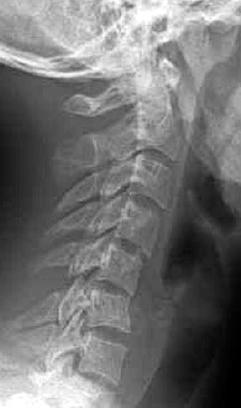
The most commonly injured, most vulnerable spinal segment following nonpenetrating trauma is the cervical spine. A critical component of the initial “assessment” of trauma patients with real or potential cervical spinal injuries is the clinical evaluation used to determine which patients require diagnostic x-ray evaluation and which do not. In a recent, comprehensive, English-language guidelines development effort, the author group used standard, clinical epidemiologic criteria for diagnostic tests to address this issue. According to protocol, the studies examined had to observe enough patients with possible injury (patients who have sustained trauma with the potential to produce spinal injury) as well as patients without injury (Fig. 7–1). The diagnostic studies had to have been conducted and compared with a gold-standard reference test indicating the presence or absence of the suspected injury.

Figure 7–1 Lateral radiograph demonstrating C5–6 ligamentous instability in an asymptomatic patient.
- Are neurologically normal
- Are not intoxicated
- Do not have neck pain or midline tenderness
- Do not have an associated injury that is distracting to the patient16–23
Based on these criteria, it has been estimated that ~14 to 58% of trauma patients evaluated in emergency rooms are asymptomatic.
The Guidelines’ author group identified nine large cohort studies that included a representative trauma population, defined symptomatic and asymptomatic patients according to the rigid criteria listed above, and reported the incidence of spinal injury in these groups of patients as detected on x-ray assessment alone or by x-ray evaluation supplemented by clinical follow-up.16–23A All nine studies were judged to provide class I medical evidence on nearly 40,000 patients in the support of a diagnostic assessment standard. Additional class II and class III medical evidence studies involving over 5000 patients were identified.24–27 This combination of medical evidence convincingly demonstrated that asymptomatic patients do not require x-ray evaluation of the cervical spine following nonpenetrating trauma. The combined negative predictive value (NPV) of cervical spine x-ray assessment of asymptomatic patients for a significant cervical spine injury as reported in the literature is virtually 100% (Table 7–3).
The Radiographic Assessment of the Cervical Spine in Symptomatic Trauma Patients
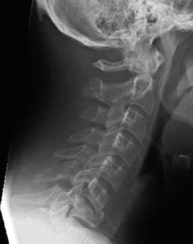
The incidence of cervical spinal injuries in the symptomatic patient population after acute trauma has been reported to be between 1.9 and 6.2%.16–20,22,23,28 The symptomatic patient is defined as any patient who does not meet all of the criteria required to be asymptomatic. Symptomatic patients may have neurological deficits related to the cervical cord or cervical roots, may have neck pain or tenderness, or may be patients in whom the designation asymptomatic is not possible (altered consciousness, intoxicated, or other distracting injury). For these patients, radiographic assessment of the cervical spine is mandatory to either identify or rule out cervical spinal injury (Fig. 7–2). The optimal radiographic assessment strategy necessary and sufficient to exclude a significant cervical spine injury in the symptomatic trauma patient has been the subject of intense scrutiny and has produced class I medical evidence on this subject, supporting the generation of standards on this topic.
| Reference | Description | Evidence Class | NPV % | PPV % |
|---|---|---|---|---|
| Hoffman et al, 200017 | Prospective study of 34,069 patients. 4309 asymptomatic. Two had clinically significant injuries All patients radiographed Note: 1 of 2 “missed injuries” did not really have a significant injury, as he was untreated and had no sequelae with clinical follow-up. The other patient developed paresthesias in his arm and was found to have a laminar fracture of C6. | I | 99.9 | 1.9 |
| Gonzales et al, 199992 | 2176 patients prospectively studied with screening examination and x-rays. One injury was detected by plain x-rays in an otherwise asymptomatic patient; however, plain x-rays missed 13 injuries overall. | I | ||
| Roth et al, 199423 | Prospective study of 682 patients admitted to emergency department with trauma: 96 were asymptomatic; none had injury. Overall incidence of injury was 2%. All patients radiographed. Follow-up clinical visit between 30 and 150 days postinjury, achieved in 43%. NPV of asymptomatic examination: 100% PPV of symptomatic examination: 2.7% | I | 100 | 2.7 |
| Lindsey et al, 199325 | 1686 patients studied retrospectively, 597 patients studied prospectively. 49 patients with cervical spine injuries were identified (overall incidence 2.1%). No patient with an injury was asymptomatic. | III | ||
| Hoffman et al, 199218 | 974 blunt trauma patients prospectively studied. Overall incidence of cervical spine injury was 2.8%. Of 353 alert, asymptomatic patients, none had a significant spine injury. Follow-up: radiographs negative in all 353. Charts, quality assurance logs, and risk management records reviewed with 3-month follow-up. NPV of asymptomatic examination: 100% PPV of symptomatic examination: 4.5% | I | 100 | 4.5 |
| Ross et al, 199222 | Prospective study of 410 patients seen at trauma center. 196 patients had asymptomatic examination; none had injury. All patients studied with plain x-rays, CT used as necessary. | I | 100 | 6.1 |
| McNamara et al, 199026 | Retrospective review of 286 patients judged to be “high risk” by mechanism of injury. 178 were asymptomatic; none had cervical spine injury. 108 were symptomatic; five had cervical spine injury. Chart follow-up performed to determine incidence of injury. NPV for asymptomatic examination: 100% PPV for symptomatic examination: 4.9% | III | 100 | 4.9 |
| Bayless and Ray, 198916 | Series of 228 patients, 211 with complete studies. Overall incidence of significant spinal injury was 1.7%. Of 122 alert, asymptomatic patients, none had a significant injury. Follow-up: x-rays negative in all 122. Charts reviewed for any subsequent referable visits within 2 years. NPV of asymptomatic examination: 100% PPV of symptomatic examination: 3% | I | 100 | 3 |
| Kreipke et al, 198919 | Prospective study of 860 patients presenting to trauma center. 324 asymptomatic; none had injury. All patients radiographed. NPV of asymptomatic examination: 100% PPV of symptomatic examination: 4% | I | 100 | 4 |
| Mirvis et al, 198927 | 408 patients studied with standard x-rays and CT. Total population seen was 4135 patients. 241 patients underwent CT because of “suspicious” x-rays, failure to visualize extremes of C-spine, or for confirmation of known fracture. Of these 241, 138 patients were clinically asymptomatic. CT served as gold standard. None of 138 patients had a clinically relevant injury (although one had a nondisplaced C7 transverse process fracture, which was treated with a collar). NPV of asymptomatic examination: 99.3–100% PPV of symptomatic examination: 12.6% | II | 99.3–100 | 12.6 |
| Neifeld et al, 198820 | Prospective study of 886 patients 244 symptomatic patients, none had injury. All patients radiographed. NPV: 100% PPV: 6.2% | I | 100 | 6.2 |
| Roberge et al, 198821 | Prospective study involving 467 trauma patients. 155 asymptomatic patients were asymptomatic, none had a spine injury. 312 were symptomatic; eight had spine injuries. All patients “scheduled to follow-up” in surgery clinic, authors state that no missed injuries have been identified. NPV of asymptomatic examination: 100% PPV of symptomatic examination: 2.5% | I | 100 | 2.5 |
| Bachulis et al, 198724 | 1823 of 4941 trauma patients studied with plain x-rays. 94 patients found to have injuries. All were symptomatic. No asymptomatic patient had a radiographically detectable injury. | III |
Abbreviations: CT, computerized tomography; NPV, negative predictive value; PPV, positive predictive value.
In a recent Guidelines’ production project on issues of cervical spine trauma, the author group identified four class I medical evidence studies, six class II studies, and 12 class III studies germane to this subject.29–49 This stringent medical evidence–based review documented class I medical evidence support for the recommendation of a standard. A three-view cervical spine x-ray series (anteroposterior, lateral, and odontoid views) of the occiput through T1, supplemented by thin-section computed tomography (CT) through areas, difficult to visualize and “suspicious” areas will detect the vast majority of cervical spinal injuries. This combination of studies represents the minimum required for clearance of the cervical spine in the symptomatic trauma patient. The negative predictive value of this combination of imaging studies is reported to be between 99 and 100% in several class II and class III medical evidence reports (Table 7–4).38,47,48,50

Figure 7–2 Lateral radiograph demonstrating C7 burst fracture in a symptomatic patient.
Stay updated, free articles. Join our Telegram channel

Full access? Get Clinical Tree


