Fluoroscopy is used to confirm adequate placement. The surgeon can also place foramen ovale electrodes through the skin into the “foramen ovale” – a hole at the base of the skull, penetrating dura mater (the tough covering of the cerebral nervous system) and lying adjacent to the mesial temporal lobe (a common site of epileptogenesis) [7]. Subdural strip electrodes are left in place for 4–7 days for seizure recording, depending on surgeon preference, and can be removed under deep conscious sedation with gentle/constant traction (no surgery required) [7].
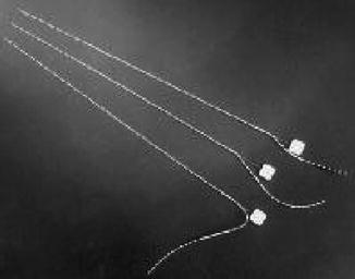
Depth electrodes are tubular, rigid or semi-rigid electrodes that penetrate brain tissue and are inserted stereotactically into the hippocampus (see image below).
These electrodes are placed with frameless or framed MRI stereotaxy using a route through the back of the head near the midline, in medical jargon an occipital, parasagittal route, so that one electrode can be placed to simultaneously record from both the amygdala and the anterior and posterior parts of the hippocampus [7]. Multimodality imaging of anatomic and angiographic studies can be combined to allow planning of trajectories to avoid vessels and improve safety of placement [7]. For instance, depth electrodes can also be placed orthogonally using an approach through the temporal bone. But blood vessels off the middle cerebral artery can travel inferiorly in the temporal lobe and be at risk of injury with this approach, so many use stereotactic angiography or magnetic resonance angiography or magnetic resonance venography (MRA/MRV). Depth electrodes can also be placed straight down, vertically with a coronal approach, but this is uncommonly used because it requires traversing the internal capsule, the nerve fibers controlling muscular function, to reach the hippocampus. Both subdural and depth electrode types are MRI-compatible [7].
In addition to defining the epileptogenic zone, the surgeon must identify functional cortex, which can be done by mapping the cortex under a grid electrode. Two electrodes in the grid are selected and stimulated with enough current to produce an effect but not so much that there are after-discharges (amount of current varies between different patients and different cortical regions) [7]. If the ictal onset zone (area where seizures initiate) is close to a crucial functional area such as motor speech, one can do an operation with the patient awake in order to map language function and to better identify the boundaries of these two (extra-operative cortical mapping is limited to an accuracy of 1 cm, which is the space between two electrodes) [7].
The most common complication of electrodes is infection −0.85 % if antibiotics are given just prior to surgery. Morbidity is highest with intracranial grids and lowest with strips. Other complications of strip electrodes include cortical contusion, cerebral edema, brain abscess, subdural empyema and subdural hematoma, placement of electrodes into brain parenchyma, accidental extraction of electrodes, superficial wound infection, and permanent neurologic deficit in <1 % [7]. Complications of grid placement are infection, transient neurologic deficit, hematoma, cerebral edema with increased intracranial pressure, and infarction. Transient neurologic deficits occur secondary to edema, hematoma or mass effect from grid, in which case the grid is removed immediately. Complications of depth electrodes are intraparenchymal hemorrhage, subarachnoid hemorrhage, vasospasm and misplacement of the electrode. The electrode can hit the brain stem or posterior cerebral artery, which can be avoided by targeting tip placement in the lateral amygdala and lateral hippocampus, making sure the hole through the occipital bone is not too close to the midline, and confirming the trajectory with an image guidance system before placing the electrode. Alternatively, the electrode can be placed in the temporal horn of the lateral ventricle (adjacent to the hippocampus) with the tip in the amygdala. The risk of permanent neurologic deficit with an occipital approach is <1 % [7].
There are many options for surgical treatment including neuroablation, neuromodulation, and surgical resection. Neuroablation can be performed via radiofrequency thermocoagulation, magnetic resonance-guided focused ultrasound surgery, laser ablation and stereotactic radiosurgery. In radiofrequency (RF) thermocoagulation, a radiofrequency generator is attached to depth electrodes that are placed deep within the brain to the presumed site of epileptogenesis. The generator creates heat by generating a current and thus induces cell death via thermocoagulation. Results are modest with only about 50 % benefiting from surgery, but this may be considered a first-line option for minimally invasive treatment [12]. Magnetic resonance-guided focused ultrasound surgery uses MRI to localize the area of interest, and then ultrasound energy is used to cause a lesion to it. Its strengths include no radiation, currently being non-invasive as no surgery is necessary (previously an opening in the skull was necessary to reduce “defocusing” caused by the skull), no trajectory restrictions and nearly real-time feedback on the lesioning effect via MR thermometry. The main drawback is inadvertent heating of the skull base resulting in injury to cranial nerves [12]. Laser ablation involves MRI-guided laser therapy that uses stereotaxy to guide thermal ablation via a laser. Advantages are that it is more precise than RF thermocoagulation, provides reliable real-time feedback and avoids skull base heating seen in ultrasound energy. The only drawback is that it is invasive, and thus incurs standard surgical risks of infection, hemorrhage, etc. [12]. Stereotactic radiosurgery focuses ionizing radiation to deep lesions with the advantage of being non-invasive but having the drawbacks of radiation (collateral tissue injury and increased risk of future malignancy) and a latent period of efficacy [12].
Within the realm of neuromodulation there is vagal nerve stimulation and deep brain stimulation. Left-sided vagal nerve stimulation (VNS) has been approved for treatment of medically refractory epilepsy as well as treatment-resistant depression, while right-sided VNS has been effective in treating heart failure. The vagus nerve is a mixed cranial nerve that is about 20 % “efferent” (in that it sends signals from the brain to the body) and 80 % “afferent” (relaying information from the body to the brain) [13]. The vagus nerve travels from the brainstem through the neck and chest and into the abdominal cavity, innervating various structures along its route. “Vagus nerve stimulation” describes any technique used to stimulate the vagus nerve – it was first discovered in the 1880s that manual massage/compression of the carotid artery in the neck could suppress seizures, theoretically by stimulating the vagus nerve. Then in the 1930s and 1940s, electrical stimulation of the vagus nerve was shown to affect brain electrical activity in cats and monkeys, and subsequent studies showed anticonvulsant effects on experimentally-induced seizures in dogs. In 1997, the FDA approved surgical implantation of a device to electrically stimulate the vagus nerve for treatment of epilepsy [13].
Related posts:


Stay updated, free articles. Join our Telegram channel

Full access? Get Clinical Tree


