and Héctor H. García3, 4
(1)
School of Medicine, Universidad Espíritu Santo, Santo, Ecuador
(2)
Department of Neurological Sciences, Hospital-Clinica Kennedy, Guayaquil, Ecuador
(3)
Cysticercosis Unit, Instituto Nacional de Ciencias Neurológicas, Lima, Peru
(4)
School of Sciences, Universidad Peruana Cayetano Heredia, Lima, Peru
Abstract
Taenia solium is one of the many species of cestodes (tapeworms) that can infect humans (Flisser 1994; Pawlowski 2002). It belongs to the phylum Platyhelminthes, class Cestoidea, order Cyclophyllidea, family Taeniidae. In general terms, tapeworms are complex organisms having complex life cycles that require at least two hosts for their completion. In the case of Taenia solium, humans are the most important definitive hosts, whereas both pigs and humans are the main intermediate hosts. The list of other animals that may act as definitive or intermediate hosts of Taenia solium is large but clinically irrelevant.
Taenia solium is one of several species of cestodes (tapeworms) that can infect humans (Flisser 1994; Pawlowski 2002). It belongs to the phylum Platyhelminthes, class Cestoidea, order Cyclophyllidea, family Taeniidae. In general terms, tapeworms are complex organisms having complex life cycles that require at least two hosts for their completion. In the case of Taenia solium, humans are the most important definitive hosts, whereas both pigs and humans are the main intermediate hosts. other animals may act as definitive or intermediate hosts of Taenia solium but their participation is clinically irrelevant.
2.1 Developmental Stages of Taenia solium
Different developmental stages of Taenia solium must be recognized, including the adult tapeworm, the egg, the embryo (oncosphere), and the larva (cysticercus). The adult T. solium has a head (scolex) that consists of four suckers and a rostellum equipped with a double crown of hooks, a narrow neck, and a large body composed by hundreds of proglottids (Fig. 2.1). The scolex of T. solium is unique in its characteristics, allowing its easy differentiation from that of T. saginata which is not armed with hooks. The body of the parasite is called the strobila and may be several meters long. Each proglottid of T. solium is equipped with a rudimentary nervous system, male and female reproductive organs, and a complex absorption and excretory system (Cárdenas-Ramirez et al. 1982). Proglottids located proximal to the scolex are immature and lack sexual organs. More sexually mature segments are located in the center of the strobila. Those mature proglottids copulate (with itself or with neighboring proglottids) and begin producing eggs. Eggs-containing proglottids are located at the distal end of the strobila. They are referred to as “gravid proglottids” and are ready to be detached from the rest of the tapeworm by a process of apolysis. Gravid proglottids contain thousands of fertile eggs which are accumulated within a long and profusely branched central uterus (Fig. 2.2).
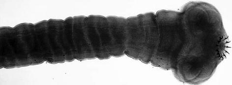
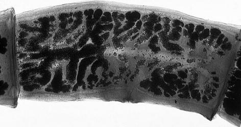
Fig. 2.1
Proglottid of Taenia solium (Image in the public domain, provider CDC/Dr. Mae Melvin)
Fig. 2.2
Scolex of Taenia solium (Image in the public domain, provider CDC/Dr. Mae Melvin)
The adult Taenia solium inhabits the small intestine of humans, where it is attached to the intestinal wall by its suckers and hooks. The parasite lives for years in the hostile intestinal environment, as it has been adapted to survive to variations in pH and to the effects of digestive enzymes. Two or three times per week, a few gravid proglottids are detached from the distal end of the body of the worm and are passed with feces. Each proglottid liberates up to 50,000 eggs which are very resistant to adverse environments; eggs can remain viable for months in water, soil, and vegetation, particularly in humid and warm environments (Aluja et al. 1987; Flisser et al. 1986).
Taenia solium eggs consist of an inner part called the oncosphere and a surrounding coat or embryophore (Fig. 2.3). The oncosphere is the parasite itself in its embryonal stage, and the embryophore is a rigid structure that protects the embryo from external environmental conditions. The latter is formed by radially distributed blocks of a keratin-like protein attached to each other by a cementing substance (Flisser 1994; Laclette et al. 1982). Once in the intestinal tract of their intermediate host (either humans or pigs), the embryophore is disrupted by enzymatic digestion. The liberated oncosphere is a solid globular organism composed of muscular, excretory, and nervous cells, as well as six embryonic hooklets—hence the term “hexacanth embryo”—and a pair of glands that are useful for migration (Pawlowski 2002). Oncospheres cross the intestinal wall and enter the bloodstream, from where they are carried to the tissues of the intermediate host where they rapidly evolve into metacestodes—the so-called postoncospheral stage—and then into larvae (cysticercus).
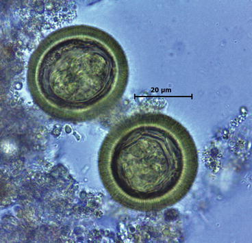
Fig. 2.3
Microscopic appearance of Taenia solium eggs (Courtesy of Juan Jimenez, MD, The Cysticercosis Working Group in Peru)
Taenia solium cysticercus is a vesicle that contains an invaginated scolex (Fig. 2.4). The vesicular wall is a membranous structure with festooned appearance composed of an outer eosinophilic layer called the cuticular mantle, a middle cellular layer with pseudoepithelial structure, and an inner layer formed by circular muscle and reticular fibers (Davis and Kornfeld 1991; Pittella 1997). The cuticular mantle is covered by microtriches which, in turn, are coated by a carbohydrate glycocalyx that increases the absorptive surface of the cysticercus. The glycocalyx is the most antigenic anatomical structure of the cysticercus. A network of excretory and neural structures is also seen inside the vesicular wall. The structure of the vesicular wall may be seen as a tegument through which adult taenias and cysticerci fulfill their metabolic and nutritional needs by absorption and diffusion (Lumsden et al. 1982; Thomas et al. 1989). The vesicular fluid is mainly composed of water but also contains calcium, glycoproteins, cholinesterase, and coproporphyrin; such composition confers the vesicular fluid fluorescent properties as well as antigenicity (Cervantes et al. 1986; Martínez-Zedillo et al. 1982).

Fig. 2.4
Macroscopic appearance of Taenia solium cysticerci. Showing vesicle and invaginated scolex (From Del Brutto et al. (1998), with permission)
The scolex located inside the vesicle has a similar structure than the adult Taenia solium, including a head armed with suckers and hooks, an elongated neck, and a rudimentary body (Fig. 2.5). Hooks are anhistous structures arranged in a double crown, with 22–28 small hooks in the outer crown, and a similar number of slightly larger hooks in the inner crown. As the larva grows, neck and body bent in a spiral within the vesicle originating the so-called spiral canal which leads from the bladder wall to the scolex. The structure of the tegument of the neck and the body of the larvae is similar to the vesicular wall, except that the cuticular mantle is thicker, and calcareous corpuscles are found in the reticular layer (Fig. 2.6).
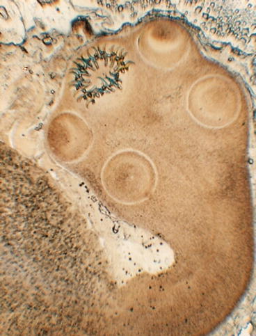
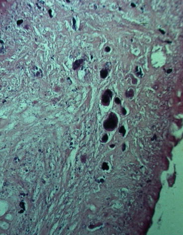
Fig. 2.5
Cysticercus of Taenia solium showing scolex with four suckers and double crown of hooks (From Del Brutto et al. (1998), with permission)
Fig. 2.6
Calcareous corpuscles in the membrane of Taenia solium cysticercus
Much of our knowledge on the different developmental stages of Taenia solium is derived from the experiments of Yoshino (1933a, b, c) who infected himself with Taenia solium cysticerci to have a constant stock of eggs to experimentally infect pigs in order to study the stages of development of embryos and metacestodes in the natural intermediate host. Yoshino observed developing metacestodes measuring 0.3 mm in the host’s tissues 6 days after oral infection. By 12 days metacestodes became cystic, and by 20–30 days, a rudimentary scolex appeared. The cysts continued to grow, measuring from 3 to 8 mm at 40–50 days. By then, cysticerci were completely formed but continued to grow for up to 1 year after infection.
2.2 Life Cycle of Taenia solium
As noted, the life cycle of Taenia solium usually involves humans and pigs (Fig. 2.7). In the normal cycle of transmission, humans are definitive hosts harboring the adult parasite in the small intestine. Gravid proglottids, detached from the distal end of the adult worm, contain infective eggs that are passed with feces of taenia carriers.