Temporal Lobe Epilepsy


Temporal Lobe Epilepsy


Introduction

Temporal lobe epilepsy (TLE) is a type of focal epilepsy where the seizures arise from the temporal lobe and adjoining structures. TLE is the most prevalent form of focal epilepsy in adults and is the syndrome that is most successfully treated by resective surgery. TLE can be further divided into mesial (mTLE) and neocortical (nTLE). The seizures are usually characterized by manual and oral automatisms (automotor seizure) as opposed to more proximal motor automatisms (hypermotor seizure) as seen predominantly in frontal lobe epilepsy, and are often preceded by a distinctive epigastric, psychic or abdominal aura. Secondary generalized tonic-clonic seizures are also not uncommon. There can be subtle differences in seizure semiology between mTLE and nTLE that will be discussed later. Lateralizing signs during seizures are important to identify and lateralize the symptomatogenic zone in TLE. It has been reported that 78% of patients presented with lateralizing signs during seizures and with a combined positive predictive value of 94%.

Epilepsy is not a static disease and is known to progress. Hippocampal sclerosis has been recognized as one of the main aetiologies in patients with mTLE. Initially, patients with TLE can be medically controlled; however, later they may evolve to develop medically intractable seizures. It is important to diagnose mTLE as early as possible, once they become medically intractable, as disabling seizures in 80-90% of patients can be eliminated by an anteriomesial temporal lobectomy. There is evidence to suggest that early resection provides the greatest opportunity for fewer psychosocial adverse events.


Anatomy of the temporal lobe

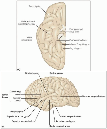
The Sylvian sulcus separates the temporal lobe from the frontal and parietal lobes with the parahippocampal gyrus marking the medial surface. There are four longitudinally orientated gyri in the temporal lobe: (1) the superior temporal gyrus (extending to the superficial transverse temporal gyrus); (2) the middle temporal gyrus; (3) the inferior temporal gyrus located on the lateral and inferolateral surface; and (4) the occipitotemporal gyrus located on the inferior surface (5.1A,B).

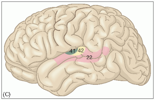
The superficial transverse temporal gyrus and adjacent portions of the superior temporal gyrus receive thalamic afferent fibres from the medial geniculate nuclei, representing the primary auditory cortex (Brodmann’s area 41 and part of 42) (5.1C). The neurons in this part of the cortex are organized tonotopically, mirroring the topographic map of frequency representation on the basilar membrane. The cortical connections for discerning the various elements of speech project to the adjacent temporal cortex, Wernicke’s area and the posterior parietal cortex, as well as other regions. Several secondary areas surround the primary auditory cortex, each with tonotopic organization.

The pulvinar nuclei and the visual cortex influence the remaining temporal gyri (known collectively as the inferior temporal association cortex). They are involved in visual and acoustic cognition, visual discrimination and pattern perception, which can be explained by their neighbouring cortices of the posterior parietal association cortex and the occipital association cortex.


Seizure semiology

Seizures of temporal lobe origin may begin with an aura. Epigastric auras are the most frequent in TLE, described often as a ‘rising sensation’ beginning in the abdomen. Other

common auras in patients with TLE include psychic, fear, auditory and olfactory auras. The occurrence of these auras gives some localization information, i.e. that they may begin in the temporal lobe but are not very helpful in lateralizing information, i.e. whether they begin in the right or left temporal lobe. Focal clonic movement has been found to lateralize the sympatogenic zone to the contralateral side of the movement, but these findings are not usually seen in the initial part of the seizure semiology in TLE. In contrast, in seizures occurring in the frontal and parietal lobes, there may be an occurrence of focal motor symptoms early in their seizure evolution.


5.1 (A) Temporal gyri and sulci from the inferior aspect. (B) Lateral aspect of left lateral lobe.


5.1 (continued) (C) Brodmann’s cortical areas of the left temporal lobe from the lateral aspect. Area 41 represents the primary auditory cortex, area 42 the associative auditory cortex and the posterior part of area 22 represents Wernicke’s area, which is responsible for the comprehension of speech.

Automatisms are a prominent feature of TLE seizures, taking many forms. Some believe unilateral motor automatisms to be of ipsilateral lateralizing value, which is most likely due to the presence of contralateral dystonic posturing masking the automatism in the contralateral limb. Oro-alimentary automatisms consisting usually of a chewing motion and lip smacking can be frequently seen in seizures of temporal lobe origin but may also occur in seizures arising outside the temporal lobe as well. Automatisms have been induced by electrical stimulation of the amygdala. Manual automatisms consist of movements involving the limbs, in which the patient appears to be fumbling, grasping, pulling at the bed sheets or manipulating an object within reach. The occurrence of motor automatisms in conjunction with dystonic posturing has been attributed to a specific primary spread of seizure discharges to subcortical brain structures and not to the seizure onset. This progression has subsequently been shown to be more common in mTLE and has been suggested as a criterion to distinguish between mTLE and nTLE. Table 5.1 lists common semiology for mTLE and nTLE, as well as noting differences.


Table 5.1 Common seizure semiology


Mesial TLE


Lateral TLE


Tripartite seizure pattern:


Tripartite seizure pattern:


aura


absence


automatism


aura


(absence less common)


automatism


Aura


Aura


visceral


visual hallucinations


cephalic


auditory hallucinations


gustatory


affective


autonomic


Partial awareness—in early stages preserved


Partial awareness—in early stages preserved


Dystonic posturing of contralateral limb with ipsilateral automatisms


Tonic posturing or jerking more common


Speech arrest during seizures with postictal dyphasia (dominant hemisphere only)


Speech arrest during seizures with postictal dyphasia (dominant hemisphere only)


Automatisms


Automatisms


oro-alimentary


oro-alimentary


gestural


gestural


sometimes prolonged


sometimes prolonged


Autonomic changes common


Autonomic changes common


Postictal confusion common


Postictal confusion common


Secondary generalization infrequent


Secondary generalization more frequent


TLE, temporal lobe epilepsy.


Table 5.2


Ictal feature


Side relative to seizure onset


Reliable signs


Unilateral dystonic posturing


Contralateral


Unilateral automatisms


Ipsilateral


Ictal speech


Non-dominant hemisphere


Postictal dysphasia


Dominant hemisphere


Forced head turning before secondary generalization (version)


Contralateral


Less reliable signs


Head turning at onset of seizure


Ipsilateral


Ictal blinking


Ipsilateral


Ictal spitting


Ipsilateral


Ictal vomitting


Non-dominant hemisphere


Postictal nose wiping


Ipsilateral

Only gold members can continue reading. Log In or Register to continue

Stay updated, free articles. Join our Telegram channel

Aug 1, 2016 | Posted by in NEUROLOGY | Comments Off on Temporal Lobe Epilepsy

Full access? Get Clinical Tree

Get Clinical Tree app for offline access