(1)
Marina Spine Center, Marina del Rey, CA, USA
1.
Place the patient in the lateral decubitus position. Approach from the convexity of the scoliosis or from the left side when possible. A left-sided approach is preferred because of ease of mobilization of the aorta compared with the vena cava. In addition, splenic retraction is easier than hepatic. Incise the skin and subcutaneous tissue from the lateral border of the paraspinous musculature over the 10th rib to the junction of the 10th rib and costal cartilage [1]. Curve the incision anteriorly from the tip of the 10th rib to the lateral rectus sheath and distally down the edge of the sheath as far as necessary for exposure (Fig. 19.1). Extend the wound slowly through each muscle layer with the electrocautery. The assistant aggressively picks up bleeders with two Ad-son forceps.
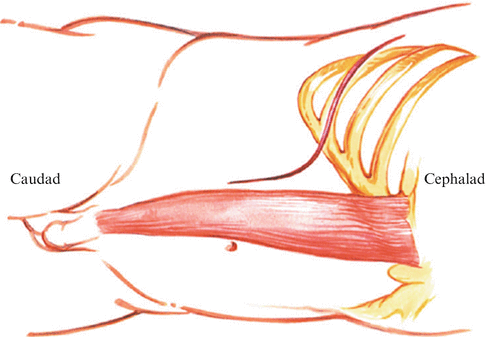

Fig. 19.1
Place the patient in a lateral decubitus position. Approach from the convexity of the scoliosis or from the left side as indicated. A left-sided-approach is preferred because of ease of mobilization of the aorta compared with the vena cava. Incise the skin and subcutaneous tissue from the lateral border of the paraspinous musculature over the 10th rib to the junction of the 10th rib and costal cartilage. Curve the incision anteriorly from the tip of the 10th rib to the lateral rectus sheath and distally down the edge of the sheath as far as necessary for exposure. Extend the wound slowly through each muscle layer to the periosteum of the 10th rib, and remove the 10th rib from the angle of the rib to the costal cartilage as in Chapter 16
2.
Open the superficial periosteum of the 10th rib to the costal cartilage. A sharp curved periosteal elevator removes the superficial and deep periosteum off the rib. Take care to avoid the neurovascular bundle on the inferior surface of the rib. Cut posteriorly at the angle of the rib and cut at the junction of rib and costal cartilage. Remove the rib. On opening the pleural space, retract the lung and open the rib bed fully with scissors (Fig. 19.2). (See Chapter 16.)
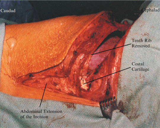

Fig. 19.2
With the rib removed, carefully delineate the costal cartilage. At this point, the intrapleural cavity is opened; the retroperitoneal cavity is still closed
3.
Split the costal cartilage with a knife along its length (Fig. 19.3). Open the undersurface of the costal cartilage and retract the two tags of cartilage [1 – 3] (Fig. 19.4).
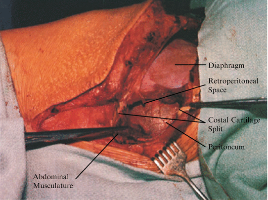
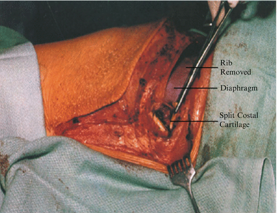

Fig. 19.3
Split the costal cartilage. Open only the most superficial layer of soft tissue under the costal cartilage enough to allow retraction of the cartilage tips

Fig. 19.4
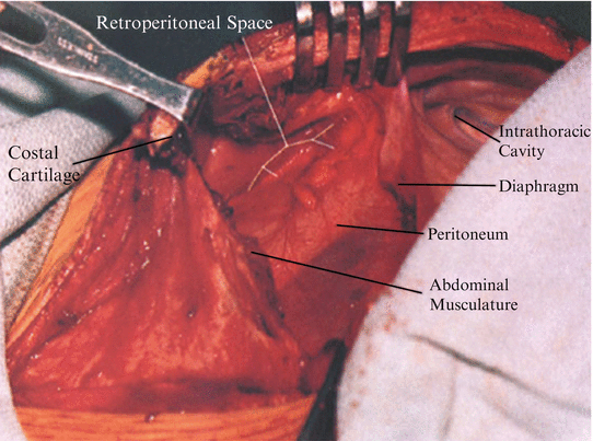
Retract the split tips of costal cartilage. Identify the insertion of the diaphragm into the cephatad cartilage tip and the insertion of the abdominal musculature into the caudad cartilage tip. Bluntly dissect under the retracted tags of cartilage and attached musculature to locate the peritoneum and retroperitoneal space. The light areolar texture of the retroperitoneal fat is the guide to the retroperitoneal space
4.
Bluntly dissect under the retracted split tips of costal cartilage to identify the retroperitoneal space (Fig. 19.5). The light areolar tissue of the retroperitoneal fat is the guide to the retroperitoneal space.

