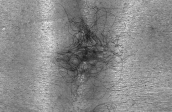
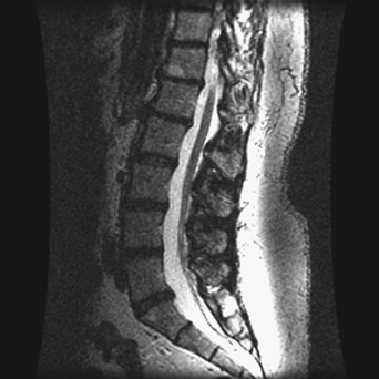
87 A 31-year-old woman reported persistent back pain a few weeks after childbirth. She had some urinary retention, lower extremity dysesthesias, and had a long-standing tuft of hair on her lower back (Fig. 87-1). A T2-weighted sagittal lumbar magnetic resonance imaging (MRI) is remarkable for a low-lying conus (Fig. 87-2). Occult spinal dysraphism FIGURE 87-1 This tuft of hair was found on the patient’s low back. A surgical de-tethering was accomplished, using neurophysiologic monitoring and microsurgical technique. The concept of spinal cord tethering has evolved to represent a gamut of congenital spinal dysraphic anomalies with a low-lying conus. In tethered cord, the conus lies below the L1 vertebral body, and the patient may or may not have neurologic deficit. The cord can be tethered primarily from a thickened filum terminale or in association with other tethering pathologies, such as a lipoma, diastematomyelia, dermal sinus tract, or myelomeningocele, and can also be iatrogenic after repair of one of the aforementioned abnormalities. Also, there have been case reports of patients who present with clinical symptoms of tethered cord with the conus above the border of the L1 vertebral body.
Tethered Cord
Presentation
Radiologic Findings
Diagnosis
Treatment
Discussion
Tethered Cord
Only gold members can continue reading. Log In or Register to continue


Full access? Get Clinical Tree


