Thalamus
Rostral to the midbrain lies the forebrain (prosencephalon, cerebrum; Fig. 1.13). The forebrain consists of the bilaterally paired diencephalon and cerebral hemisphere on each side and is by far the largest derivative of the three basic embryological divisions of the brain. The diencephalon is continuous with the rostral part of the midbrain and lies between the brainstem and the cerebral hemisphere. From dorsal to ventral, the diencephalon is comprised of the epithalamus, thalamus, subthalamus and hypothalamus, of which the thalamus is the largest. The thalamus consists of numerous nuclei, most of which have extensive reciprocal connections with the cerebral cortex. Of particular note are:
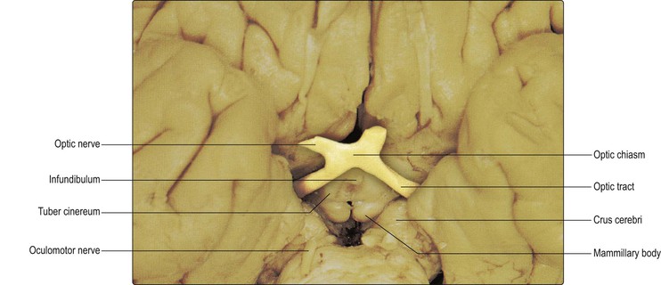
The diencephalon is almost entirely surrounded by the cerebral hemisphere; consequently, little of its structure can be seen externally, apart from the ventral portion of the hypothalamus, which can be seen on the base of the brain (Fig. 12.1). Immediately caudal to the optic chiasm is a small midline elevation, the tuber cinereum. From its apex extends the infundibulum or pituitary stalk, which attaches to the pituitary gland. Caudal to the tuber cinereum, a pair of rounded eminences, the mammillary bodies, are located on either side of the midline. These contain the mammillary nuclei of the hypothalamus. The hypothalamus lies below the thalamus, extending medial and ventral to the subthalamus. It has important connections with the limbic system, a controlling influence upon the activity of the autonomic nervous system and a central role in neuroendocrine function, partly through its relationship with the pituitary gland. The hypothalamus is discussed further in Chapter 16.
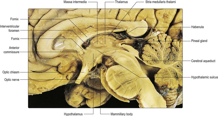
The other parts of the diencephalon can be seen in sagittal and coronal sections of the brain (Figs 12.2, 12.3). The diencephalon forms the lateral wall of the third ventricle. The dorsal part of the ventricular wall is formed by the thalamus and the ventral part by the hypothalamus. The epithalamus is a relatively small part of the diencephalon located in its most caudal and dorsal region, immediately rostral to the superior colliculus of the midbrain. It consists principally of the pineal gland and the habenula (habenular nuclei). The pineal gland is an endocrine organ. It synthesises the hormone melatonin. The pineal gland has been implicated in control of the sleep/waking cycle (circadian rhythm) and in regulation of the onset of puberty. The habenular nuclei have connections with the limbic system (Ch. 16).
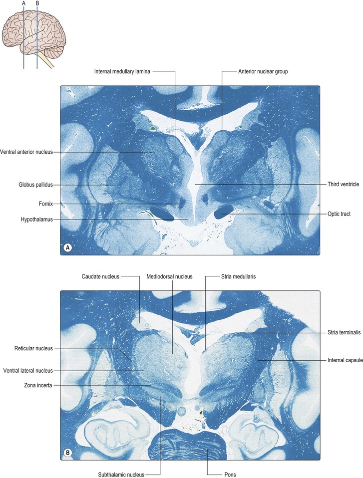
The subthalamus lies beneath the thalamus and dorsolateral to the hypothalamus, with its ventrolateral aspect against the internal capsule. It contains two notable cell groups, the subthalamic nucleus and the zona incerta. The subthalamic nucleus is located in the ventrolateral part of the subthalamus, immediately medial to the internal capsule. It has the shape of a biconvex lens in coronal section. The subthalamic nucleus has prominent connections with the globus pallidus and the substantia nigra and is important in the control of movement. It is discussed in more detail in Chapter 14. The zona incerta is a rostral extension of the brainstem reticular formation. Several important fibre systems traverse the subthalamus en route to the thalamus. These include ascending sensory projections (medial lemniscus, spinothalamic tracts, trigeminothalamic tracts), cerebellothalamic fibres from the dentate nucleus and pallidothalamic fibres from the internal segment of the globus pallidus. The latter group of fibres envelop the zona incerta as the lenticular fasciculus and thalamic fasciculus (see Fig. 14.8).
Topographical anatomy of the thalamus
External features
The thalamus has been likened in size and shape to a small hen’s egg. Together with the hypothalamus, it forms the lateral wall of the third ventricle, the transition between the two being marked by a faint groove, the hypothalamic sulcus. In most individuals, the two thalami are joined across the thin slit of the ventricle by the interthalamic adhesion or massa intermedia. A fascicle of nerve fibres, the stria medullaris, which has limbic connections, courses along the dorsomedial margin of the thalamus (Fig. 12.2). Along this line, the ependymal lining of the third ventricle spans the narrow lumen to form the ventricular roof.
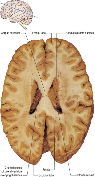
The anterior pole of the thalamus extends as far as the interventricular foramen, through which the third and lateral ventricles are in continuity. Lateral to the thalamus lies the posterior limb of the internal capsule and anterolateral lies the head of the caudate nucleus (Fig. 12.4). The dorsal aspect of the thalamus thus forms part of the floor of the body of the lateral ventricle. Another fascicle of nerve fibres with limbic connections, the stria terminalis, marks the boundary between thalamus and caudate (Fig. 12.4). Ventral to the thalamus lie the subthalamus and hypothalamus; caudal to it lies the midbrain.
Related posts:


Stay updated, free articles. Join our Telegram channel

Full access? Get Clinical Tree


