The Auditory System: Introduction
Anatomy and Function
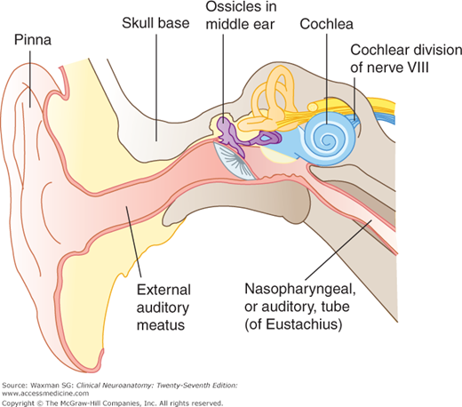
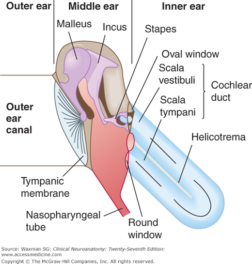
The cochlea, within the inner ear, is the specialized organ that registers and transduces sound waves. It lies within the cochlear duct, a portion of the membranous labyrinth within the temporal bone of the skull base (Fig 16–1; see also Chapter 11). Sound waves converge through the pinna and outer ear canal to strike the tympanic membrane (Figs 16–1 and 16–2). The vibrations of this membrane are transmitted by way of three ossicles (malleus, incus, and stapes) in the middle ear to the oval window, where the sound waves are transmitted to the cochlear duct.
Two small muscles can affect the strength of the auditory signal: the tensor tympani, which attaches to the eardrum, and the stapedius muscle, which attaches to the stapes. These muscles may dampen the signal; they also help prevent damage to the ear from very loud noises.
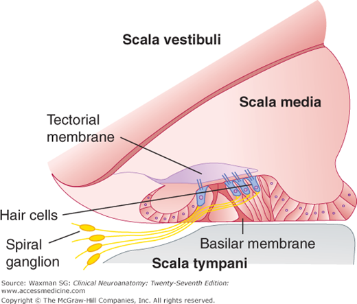
The inner ear contains the organ of Corti within the cochlear duct (Fig 16–3). As a result of movement of the stapes and tympanic membrane, a traveling wave is set up in the perilymph within the scala vestibuli of the cochlea. The traveling waves propagate along the cochlea; high-frequency sound stimuli elicit waves that reach their maximum near the base of the cochlea (ie, near the oval window). Low-frequency sounds elicit waves that reach their peak, in contrast, near the apex of the cochlea (ie, close to the round window). Thus, sounds of different frequencies tend to excite hair cells in different parts of the cochlea, which is tonotopically organized.
The human cochlea contains more than 15,000 hair cells. These specialized receptor cells transduce mechanical (auditory) stimuli into electrical signals.
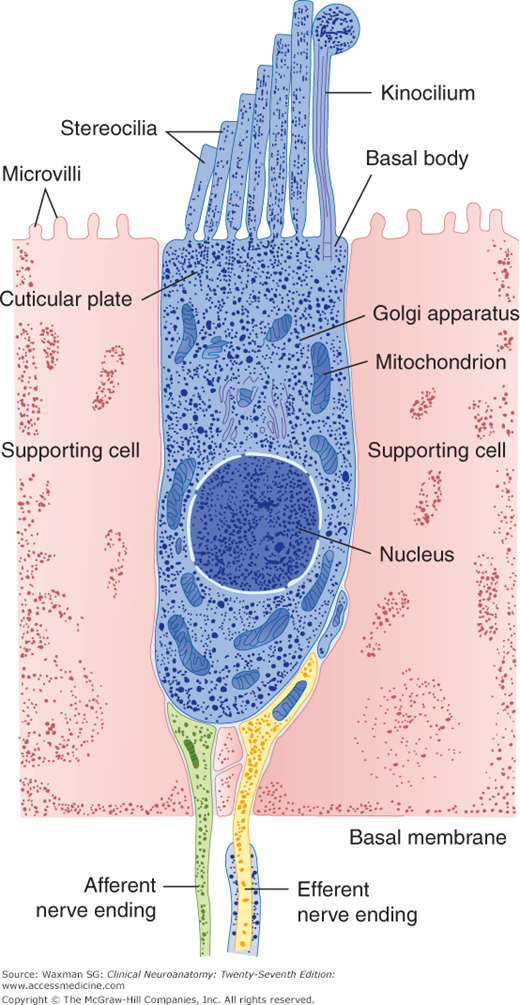
The traveling waves within the perilymph stimulate the organ of Corti through the vibrations of the tectorial membrane against the kinocilia of the hair cells (Figs 16–3 and 16–4). The mechanical distortions of the kinocilium of each hair cell are transformed into depolarizations, which open calcium channels within the hair cells. These channels are clustered close to synaptic zones. Influx of calcium, after opening of these channels, evokes release of neurotransmitter, which elicits a depolarization in peripheral branches of neurons of the cochlear ganglion. As a result, action potentials are produced that are transmitted to the brain along axons that run within the cochlear nerve.
Figure 16–4
Structure of hair cell. (Reproduced with permission from Hudspeth AJ: The hair cells of the inner ear. They are exquisitely sensitive transducers that in human beings mediate the senses of hearing and balance. A tiny force applied to the top of the cell produces an electrical signal at the bottom, Sci Am Jan;248(1):54–64, 1983.)


